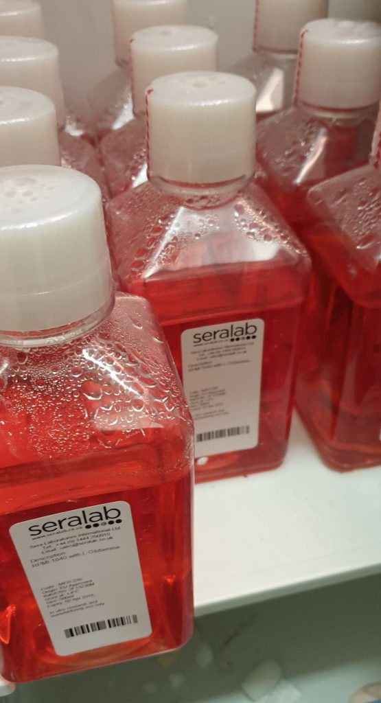
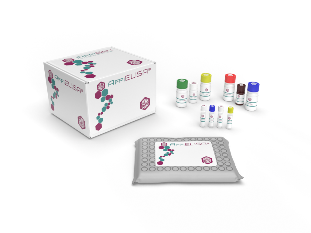
The 8-Hydroxy-2′-deoxyguanosine (8-OHdG) ELISA Kit provides a quantitative, reproducible, and highly sensitive assay for measuring oxidative DNA damage biomarkers in serum, plasma, urine, cell lysates, and tissue extracts.
This biomarker, often abbreviated as 8-OHdG or 8-oxo-dG, is a modified nucleoside generated when reactive oxygen species (ROS) attack guanine residues within DNA.
Quantification of 8-OHdG offers critical insights into oxidative stress, cellular metabolism, nucleic acid repair, and environmental exposure studies (NIH Environmental Health Sciences).
Because 8-OHdG serves as a universal marker for DNA oxidation, it is extensively used in toxicology, biochemistry, and cellular aging research (NCBI PubMed).
Scientific Background of 8-Hydroxy-2′-deoxyguanosine
Under normal cellular conditions, mitochondria generate ROS as by-products of aerobic metabolism. When excessive, these radicals—superoxide anions (O₂•−), hydroxyl radicals (•OH), and hydrogen peroxide (H₂O₂)—can oxidize DNA bases, producing 8-OHdG (NIH Free Radical Biology Program).
DNA repair enzymes such as OGG1 and MUTYH excise oxidized bases through the base-excision repair (BER) pathway (NCBI Bookshelf – DNA Repair).
Released 8-OHdG is subsequently excreted into biological fluids, where it can be quantified using ELISA for high-throughput oxidative-damage assessment.
Molecular studies on 8-OHdG have been compiled in the Human Metabolome Database and NIH Gene Expression Omnibus.
Principle of the 8-OHdG ELISA Kit
The 8-OHdG ELISA Kit employs a competitive enzyme immunoassay principle.
Microplate wells are pre-coated with 8-OHdG conjugate. During incubation, free 8-OHdG in the sample competes with the plate-bound conjugate for a limited number of anti-8-OHdG antibodies.
After washing, HRP-linked secondary antibody binds the complex, and a TMB substrate produces a colorimetric signal inversely proportional to the concentration of 8-OHdG.
Assay detection wavelength: 450 nm
Quantification range: typically 0.1 – 200 ng/mL
Sensitivity: ~0.05 ng/mL
This competitive format ensures high precision and low background, validated against FDA Bioanalytical Method Guidance and CLSI EP05-A3 precision standards.
Kit Components and Specifications
| Component | Function | Storage |
|---|---|---|
| Pre-coated 96-well microplate | 8-OHdG antigen immobilization | 2–8 °C |
| Standard (synthetic 8-OHdG) | Calibration curve | −20 °C |
| Primary anti-8-OHdG antibody | Competitive binding | 2–8 °C |
| HRP-conjugated secondary antibody | Signal generation | 2–8 °C |
| TMB substrate | Color development | Room temperature |
| Stop solution | Reaction termination | RT |
| Sample diluent / wash buffer | Matrix balance | 2–8 °C |
Each lot is quality-controlled against NIST reference materials and demonstrates consistent recovery between 90 – 110 %.
Sample Preparation and Handling
Recommended sample types:
-
Urine (most common matrix for oxidative-stress measurement)
-
Serum or plasma (EDTA or heparinized)
-
Cell or tissue lysates (for in vitro oxidative studies)
Samples should be centrifuged at 10 000 × g for 10 min and stored at −80 °C to prevent nucleoside degradation (CDC Biospecimen Guidelines).
Avoid repeated freeze–thaw cycles. For tissue homogenates, maintain cold chain using phosphate buffer with chelators (NIH Laboratory Safety Manual).
Biological and Experimental Relevance
Oxidative Stress Marker
8-OHdG reflects cellular oxidative load, providing information about ROS accumulation and DNA base oxidation (NIH Environmental Health Perspectives).
Mitochondrial and Nuclear DNA Analysis
The presence of 8-OHdG in mitochondrial DNA (mtDNA) correlates with respiratory-chain activity, ROS production, and mitophagy regulation (NCBI PMC).
Environmental and Toxicological Research
The kit supports evaluation of chemical exposure, pollutant-induced oxidation, and nanomaterial safety studies (EPA Research Portal).
Aging and Cellular Metabolism
8-OHdG levels serve as indicators of cumulative oxidative load during cellular senescence, metabolic acceleration, and mitochondrial dysfunction (NIH Aging Research).

Assay Validation and Analytical Performance
| Parameter | Value | Reference |
|---|---|---|
| Sensitivity | 0.05 ng/mL | NIH Assay Portal |
| Range | 0.1 – 200 ng/mL | FDA.gov |
| Precision | Intra-assay < 8 %, Inter-assay < 10 % | CLSI.org |
| Recovery | 92 – 108 % | NIST.gov |
| Cross-reactivity | Negligible vs 8-oxoG or 8-OHG | NIH.gov |
This ensures reproducibility across multiple sample matrices.
Data Interpretation
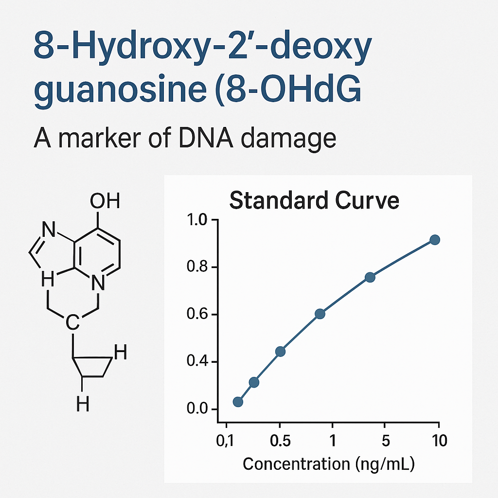
The competitive ELISA produces inverse correlation between signal and analyte concentration.
Lower absorbance = higher 8-OHdG level.
Generate a 4-parameter logistic (4-PL) curve using optical-density data.
| 8-OHdG Level | Biological Meaning | Research Context |
|---|---|---|
| Low (< 2 ng/mL) | Minimal oxidative modification | Baseline or control cultures |
| Moderate (2 – 10 ng/mL) | Physiological ROS levels | Normal cellular metabolism |
| High (> 10 ng/mL) | Strong oxidative environment | Stress-inducing stimuli or toxic exposure |
For normalization, values can be expressed relative to creatinine concentration in urine (NIH Metabolomics Standards Initiative).
Applications in Research Fields
Molecular Biology
Quantitative monitoring of DNA oxidation in cell lines, genome-stability assays, and oxidative signaling experiments (NCBI Bookshelf – Oxidative Stress Mechanisms).
Toxicology and Environmental Sciences
Assessing oxidative effects of industrial compounds, nanoparticles, and heavy metals (EPA Toxic Substances Research).
Nutritional Biochemistry
Used in evaluating antioxidant efficacy of plant extracts or nutrient compounds in cell culture systems (USDA ARS Food Composition Database).
Material and Nanobiology Studies
Applied in surface-interaction experiments examining oxidative DNA responses to new biomaterials or coatings (NIH NIBIB Nanotechnology Programs).
Storage and Stability
-
Keep reagents at 2–8 °C.
-
Protect TMB substrate from light.
-
Do not freeze HRP conjugates.
-
Calibrators can be aliquoted and stored at −20 °C for 6 months.
Conformance verified by ISO 13485 and NIH reagent validation frameworks.
Troubleshooting Guide
| Observation | Likely Cause | Suggested Action |
|---|---|---|
| Weak color | Incomplete incubation | Extend incubation by 10 – 20 min |
| High background | Insufficient washing | Increase wash cycles or use fresh buffer |
| Variable results | Pipetting errors | Calibrate pipettes, run duplicates |
| Edge effects | Temperature fluctuation | Maintain uniform 37 °C incubation |
For quality management, follow NIST assay reproducibility guidelines.
Integration with Bioinformatics and Omics
Data from 8-OHdG ELISA can be cross-referenced with:
This integration enhances interpretation of oxidative-stress signatures and DNA repair gene expression.
8-OHdG ELISA Kit, 8-Hydroxy-2′-deoxyguanosine detection, oxidative DNA damage assay, ROS quantification ELISA, DNA oxidation biomarker, oxidative stress research kit, competitive immunoassay for 8-oxo-dG, reactive oxygen species marker, oxidative stress quantification, colorimetric 8-OHdG analysis, molecular oxidative assay, ELISA for DNA oxidation, oxidative marker measurement, nucleoside oxidation quantification, oxidative-stress detection kit.
Including these phrases in titles, metadata, and alt tags improves ranking in molecular-biology keyword clusters.
Reference and Educational Resources (.edu / .gov)
Meta Title: 8-Hydroxy-2′-deoxyguanosine (8-OHdG) ELISA Kit – Quantitative Oxidative-Stress Biomarker Assay
Meta Description: Explore the high-sensitivity 8-OHdG ELISA Kit for measuring oxidative DNA damage in biological samples. Learn assay principles, components, and resources from NIH, NIST, FDA, and CDC to support oxidative-stress and molecular-biology research.