High-Fidelity PCR: Advanced Principles, Applications, and Optimization for Precision Molecular Biology

High-Fidelity PCR has become a core molecular biology technique in laboratories where sequence accuracy, reproducibility, and downstream compatibility are essential. As experimental pipelines increasingly rely on cloning, next-generation sequencing (NGS), synthetic biology, and genome engineering, conventional PCR enzymes no longer meet the accuracy requirements demanded by modern workflows.

Unlike routine PCR—where yield and speed may be prioritized—High-Fidelity PCR emphasizes correctness. Even a single base substitution introduced during amplification can compromise cloning efficiency, alter protein function, or generate misleading sequencing data. This makes high-fidelity amplification a quality-control step, not merely an amplification step.

For foundational PCR concepts, the National Center for Biotechnology Information (NCBI) provides a comprehensive overview:
https://www.ncbi.nlm.nih.gov/probe/docs/techpcr/
https://www.ncbi.nlm.nih.gov/books/NBK589663/

AffiPCR® EVO HS Super-Fidelity DNA Polymerase

Molecular Basis of Fidelity in DNA Polymerases

Proofreading Exonuclease Activity

The defining feature of high-fidelity DNA polymerases is 3′→5′ exonuclease proofreading activity. During nucleotide incorporation, if an incorrect base is added, the polymerase pauses, excises the mismatched nucleotide, and resumes synthesis with the correct base.

This mechanism dramatically reduces:

  • Base substitutions

  • Frameshift mutations

  • Accumulated sequence drift across cycles

A detailed biochemical discussion of proofreading activity can be found in this NIH-hosted review:
https://pmc.ncbi.nlm.nih.gov/articles/PMC2685094/

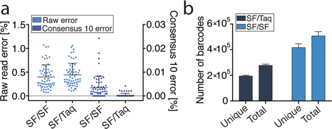
Error Rate Comparison

Polymerase Type Approximate Error Rate
Standard Taq ~1 × 10⁻⁴ – 10⁻⁵
High-Fidelity ~1 × 10⁻⁶ – 10⁻⁷

Classic fidelity measurements and experimental comparisons are discussed in PubMed literature:
https://pubmed.ncbi.nlm.nih.gov/1842916/

This order-of-magnitude difference is critical when amplifying long fragments or templates destined for sequencing.

Why High-Fidelity PCR Is Essential in Modern Workflows
https://media.springernature.com/lw685/springer-static/image/art%3A10.1038%2Fs41598-019-39762-6/MediaObjects/41598_2019_39762_Fig3_HTML.png?utm_source=chatgpt.com

1. Gene Cloning and Expression Vector Construction

When PCR products are cloned into plasmids:

  • Any nucleotide error becomes a permanent part of the construct

  • Screening time increases dramatically

  • Protein expression may be altered or abolished

Many university cloning manuals explicitly recommend proofreading polymerases for insert generation:
https://yen.fccc.edu/docs/pentr_dtopo_man.pdf
https://openwetware.org/wiki/Gibson_Assembly

Next-Generation Sequencing (NGS) Library Preparation

PCR errors introduced before sequencing:

  • Appear as false variants

  • Inflate diversity estimates

  • Reduce confidence in low-frequency mutation detection

NIH-hosted resources on PCR bias and sequencing artifacts highlight the importance of enzyme choice:
https://www.ncbi.nlm.nih.gov/pmc/articles/PMC5428825/
https://www.ncbi.nlm.nih.gov/pmc/articles/PMC6517913/

Long-Range and GC-Rich Template Amplification

High-fidelity PCR systems are widely used for:

  • Long genomic regions (>5 kb)

  • GC-rich promoters

  • Repetitive or structured DNA

University protocol references include:
https://web.as.uky.edu/biology/faculty/mirabito/BIO%20510%20Fall%202007/Expand%20Long%20Template.pdf
https://openwetware.org/wiki/Long-range_PCR

Site-Directed Mutagenesis and Synthetic Biology

When introducing intentional mutations, background errors are undesirable. High-fidelity PCR ensures:

  • Only the designed mutation is present

  • Reduced off-target sequence noise

  • Higher success rates in downstream validation

Educational mutagenesis resources:
https://www.addgene.org/protocols/quickchange-mutagenesis/
https://openwetware.org/wiki/Site-directed_mutagenesis

Primer Design: A Major Determinant of High-Fidelity Success

https://upload.wikimedia.org/wikipedia/commons/thumb/1/19/Primer_dimers_formation_En.PNG/500px-Primer_dimers_formation_En.PNG?utm_source=chatgpt.com

Even the best polymerase cannot compensate for poor primer design.

Key Design Parameters (University-Backed)

  • Length: 18–24 nucleotides

  • GC content: 40–60%

  • Minimal secondary structure

  • Balanced melting temperatures (Tm)

Authoritative academic primer design resources:

Cycling Conditions Optimized for High-Fidelity PCR

Critical Parameters

Parameter Recommendation
Annealing temperature Use calculated Tm, often higher than standard PCR
Extension time Match polymerase speed and fragment length
Cycle number Minimize (typically 25–30 cycles)
Hot-start Strongly recommended

Educational PCR optimization guides:

Common High-Fidelity PCR Pitfalls and Solutions

Nonspecific Amplification

  • Increase annealing temperature

  • Reduce primer concentration

  • Redesign primers with improved specificity

Low Yield

  • Extend elongation time

  • Increase template quality

  • Verify Mg²⁺ concentration

Primer Dimers

  • Avoid 3′ complementarity

  • Use hot-start protocols

  • Redesign primers if persistent

Troubleshooting references:
https://openwetware.org/wiki/PCR_troubleshooting
https://www.ncbi.nlm.nih.gov/pmc/articles/PMC4213345/

Quality Control and Standardization in PCR Workflows

High-fidelity PCR is commonly integrated into validated research pipelines, where consistency matters.

Recommended controls:

  • No-template controls (NTC)

  • Positive amplification controls

  • Replicate reactions

  • Reference materials

NIST provides DNA reference materials used in PCR-based assays:

These resources highlight how PCR accuracy is central to reproducibility in molecular biology.

Where High-Fidelity PCR Delivers the Most Value

High-Fidelity PCR is particularly suited for:

  • Cloning and plasmid construction

  • Amplicon sequencing

  • Synthetic biology

  • CRISPR donor preparation

  • Long-range PCR

  • Variant confirmation

It is not simply a premium alternative—it is a risk-reduction strategy for experiments where errors are costly.

Product Integration: High-Fidelity PCR for Reliable, Sequence-Accurate Amplification

A dedicated High-Fidelity PCR solution supports laboratories seeking:

  • Reduced mutation rates

  • Cleaner amplification profiles

  • Faster downstream validation

  • Improved reproducibility

By combining proofreading polymerase activity with optimized buffer systems, High-Fidelity PCR enables sequence-ready DNA amplification that integrates seamlessly into cloning, sequencing, and molecular engineering workflows.
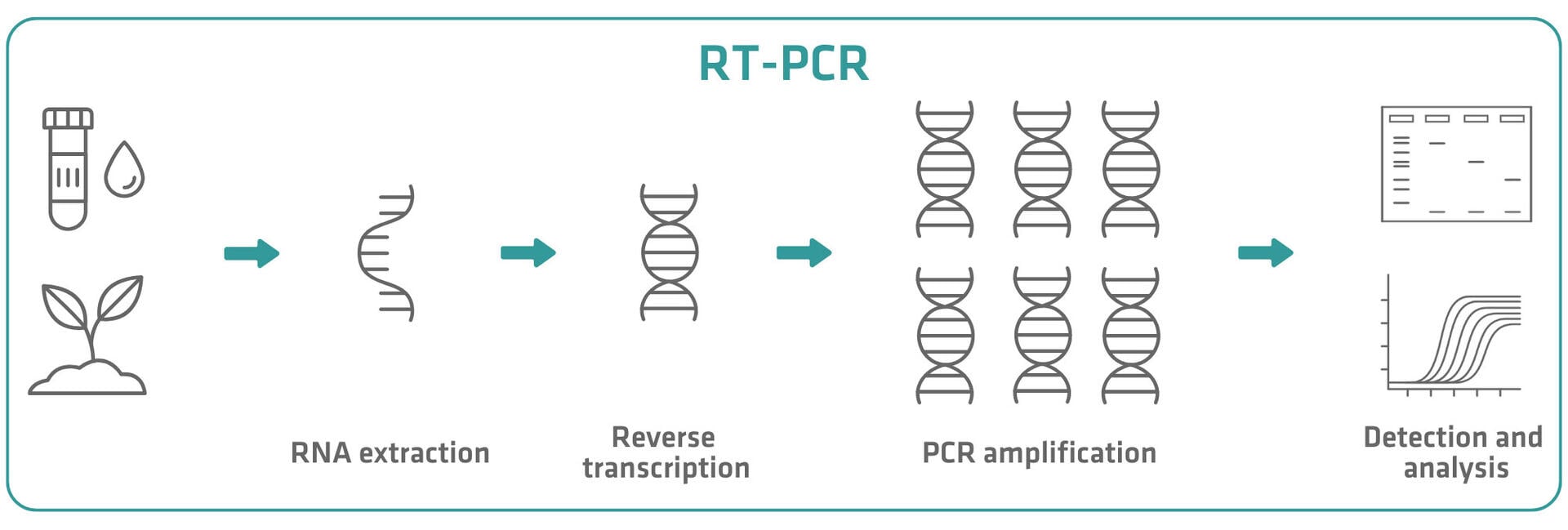
Conventional RT-PCR and Reverse Transcriptase: A Comprehensive Laboratory Guide to RNA Analysis

Conventional Reverse Transcription Polymerase Chain Reaction (RT-PCR) is a foundational molecular biology technique used to analyze RNA expression, transcript presence, and RNA-derived genetic material. By combining reverse transcription with endpoint PCR amplification, conventional RT-PCR enables laboratories to convert RNA into complementary DNA (cDNA) and subsequently detect specific sequences using standard PCR workflows.

Despite the emergence of real-time RT-PCR and sequencing-based transcriptomics, conventional RT-PCR remains indispensable in research, biotechnology, education, and method development laboratories due to its simplicity, flexibility, and robustness.

This article provides an in-depth, laboratory-oriented overview of conventional RT-PCR, focusing on reverse transcriptase enzymes, workflow design, optimization strategies, and practical applications, before introducing how a Conventional RT-PCR Kit / Reverse Transcriptase integrates into routine molecular biology workflows.

RNA analysis as a cornerstone of molecular biology

RNA serves as the functional intermediary between genotype and phenotype, reflecting gene expression, regulation, and cellular response. Studying RNA allows researchers to:

  • detect gene transcription

  • analyze alternative splicing

  • monitor expression changes

  • study RNA viruses and transcripts

Educational overviews from NIH and Genome.gov explain the central role of RNA in molecular biology:
https://www.ncbi.nlm.nih.gov/books/NBK26850/
https://www.genome.gov/genetics-glossary/RNA

Because PCR enzymes amplify DNA—not RNA—reverse transcription is required to convert RNA into a DNA template suitable for amplification.

The essential guide to RT-PCR | INTEGRA

What is conventional RT-PCR?

Conventional RT-PCR is a two-step molecular technique consisting of:

  1. Reverse transcription (RT) – synthesis of cDNA from RNA

  2. Conventional PCR – amplification of the cDNA using sequence-specific primers

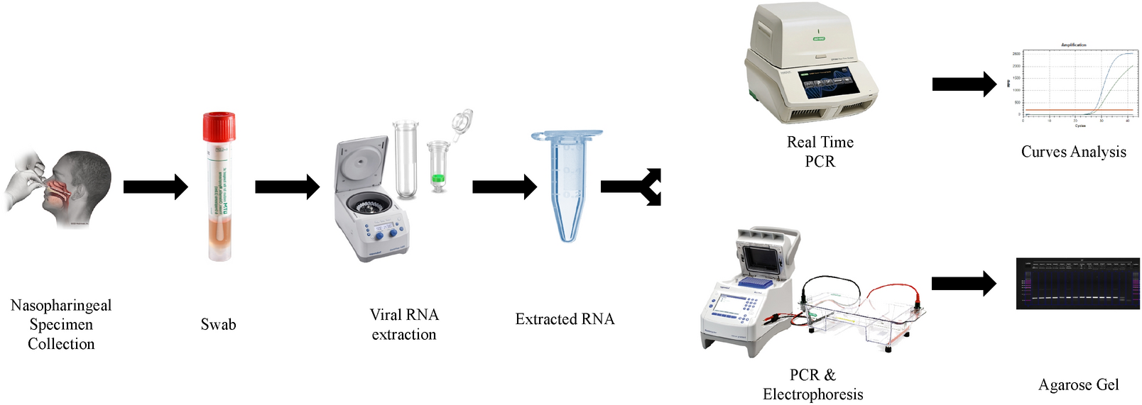
The final PCR product is detected at the endpoint, most commonly by agarose gel electrophoresis.

NIH-hosted molecular biology resources describe RT-PCR principles and workflows in detail:
https://www.ncbi.nlm.nih.gov/books/NBK55396/
https://www.ncbi.nlm.nih.gov/pmc/articles/PMC7141722/

Reverse transcriptase: the key enzymatic component

 Origin and function of reverse transcriptase

Reverse transcriptase (RT) enzymes were originally identified in retroviruses, where they catalyze the synthesis of DNA from an RNA template. In molecular biology laboratories, engineered and purified reverse transcriptases are used to generate cDNA for downstream applications.

Educational explanations of reverse transcriptase function are available from NIH and NCBI:
https://www.ncbi.nlm.nih.gov/books/NBK26852/
https://www.ncbi.nlm.nih.gov/pmc/articles/PMC3058489/

 Common properties of laboratory reverse transcriptases

Laboratory-grade reverse transcriptases are typically characterized by:

  • RNA-dependent DNA polymerase activity

  • reduced RNase H activity (to preserve RNA template)

  • compatibility with a range of primers

  • activity across moderate temperature ranges

These properties influence cDNA yield, length, and representation.

The Basics of Reverse Transcription PCR (RT-PCR)

RT-PCR workflow: step-by-step laboratory process

Step 1: RNA isolation

High-quality RNA is essential for reliable RT-PCR results. RNA integrity, purity, and absence of inhibitors directly impact reverse transcription efficiency.

NIH and academic protocols for RNA extraction emphasize RNase control and sample handling:
https://www.ncbi.nlm.nih.gov/pmc/articles/PMC5452224/
https://www.ncbi.nlm.nih.gov/books/NBK55416/

Step 2: Reverse transcription (cDNA synthesis)

RNA is converted into cDNA using reverse transcriptase, primers, dNTPs, and buffer. Reaction conditions depend on:

  • RNA template type (total RNA, mRNA, viral RNA)

  • primer strategy

  • enzyme characteristics

University molecular biology manuals describe RT reaction setup and optimization:
https://open.oregonstate.education/microbiology/chapter/rt-pcr/

Step 3: Conventional PCR amplification

The synthesized cDNA is amplified using standard PCR reagents and thermal cycling.
This step follows the same denaturation–annealing–extension logic as conventional PCR.

https://www.ncbi.nlm.nih.gov/pmc/articles/PMC2836090/

Step 4: Endpoint detection

Amplified products are analyzed by agarose gel electrophoresis to confirm:

  • presence or absence of transcripts

  • expected amplicon size

  • reaction specificity

https://www.ncbi.nlm.nih.gov/books/NBK55416/

Primer strategies for reverse transcription

Primer choice during reverse transcription strongly influences which RNA molecules are converted into cDNA.

Common primer types:

  • Oligo(dT) primers – target poly(A)+ mRNA

  • Random primers – generate broad cDNA coverage

  • Gene-specific primers – focus on a single transcript

Educational comparisons of RT priming strategies are provided by NIH-hosted resources:
https://www.ncbi.nlm.nih.gov/pmc/articles/PMC4478221/

Primer selection affects sensitivity, specificity, and transcript representation.

Optimization considerations in conventional RT-PCR

Successful RT-PCR requires optimization at both RT and PCR stages.

Key parameters include:

  • RNA input amount

  • RT incubation temperature and duration

  • primer concentration

  • Mg²⁺ concentration

  • PCR annealing temperature

Academic guides emphasize performing controls to distinguish RT failure from PCR failure:
https://www.ncbi.nlm.nih.gov/books/NBK55396/

Controls and quality assurance in RT-PCR

Standard laboratory controls include:

  • No-RT controls (to detect genomic DNA contamination)

  • No-template controls (NTC)

  • Positive RT controls

CDC molecular biology training materials illustrate control strategies relevant to RT-PCR workflows:
https://www.cdc.gov/labtraining/training-courses/pcr.html

Including appropriate controls is essential for result interpretation and reproducibility.

Applications of conventional RT-PCR in laboratories

 Gene expression screening

RT-PCR is commonly used to assess whether a gene is transcriptionally active in a sample.
https://www.ncbi.nlm.nih.gov/pmc/articles/PMC7141722/

 RNA virus research (research use)

Conventional RT-PCR enables qualitative detection of RNA viruses in research and environmental studies.
https://www.ncbi.nlm.nih.gov/pmc/articles/PMC5452224/

 Splice variant and transcript structure analysis

RT-PCR allows detection of alternative splicing events by amplifying across exon junctions.
https://www.ncbi.nlm.nih.gov/pmc/articles/PMC5868358/

 Method development and assay validation

Conventional RT-PCR is often used as a developmental step before implementing quantitative RT-PCR or sequencing assays.

Limitations of conventional RT-PCR

While powerful, conventional RT-PCR has inherent limitations:

  • endpoint (non-quantitative) analysis

  • gel-based detection

  • limited dynamic range

  • sensitivity to RNA degradation

Comparative reviews discuss where conventional RT-PCR fits relative to qRT-PCR and RNA-Seq:
https://www.ncbi.nlm.nih.gov/pmc/articles/PMC7141722/

  • conventional RT-PCR

  • reverse transcriptase enzyme

  • RNA to cDNA synthesis

  • endpoint RT-PCR analysis

  • reverse transcription PCR kit

  • transcript detection laboratory

Practical value of a Conventional RT-PCR Kit / Reverse Transcriptase

A Conventional RT-PCR Kit or Reverse Transcriptase supports laboratories by providing:

  • reliable RNA-to-cDNA conversion

  • compatibility with standard PCR systems

  • flexibility in primer selection

  • suitability for research and method development

Such products are widely used in academic research, biotechnology R&D, training laboratories, and quality control method development, where simplicity and reproducibility are essential.

Authoritative educational references (.edu / .gov)

Conventional PCR: A Comprehensive Laboratory Guide to Principles, Optimization, and Applications

Conventional Polymerase Chain Reaction (PCR) remains one of the most fundamental and indispensable molecular biology techniques in use today. While advanced technologies such as real-time PCR, digital PCR, and next-generation sequencing continue to expand analytical capabilities, conventional PCR persists as a core laboratory method for DNA amplification, screening, verification, and method development.

This extended guide explores conventional PCR in depth, covering its molecular principles, reaction design, optimization strategies, laboratory workflows, and diverse applications, with a strong focus on practical laboratory use rather than clinical interpretation.

Historical context and scientific foundation of PCR

PCR was first described in the 1980s and rapidly transformed molecular biology by enabling in vitro amplification of specific DNA sequences. Its development eliminated the need for cloning DNA fragments into living cells for amplification, significantly accelerating genetic analysis.

Educational overviews from U.S. government and academic institutions explain the historical and scientific importance of PCR:

Conventional PCR laid the foundation for nearly all modern nucleic acid–based techniques used today.

AffiPCR® Green Taq Mix

Molecular mechanism of conventional PCR

At its core, conventional PCR relies on enzymatic DNA replication driven by temperature cycling. The reaction mimics natural DNA synthesis but does so in a controlled, cyclic manner.

 Denaturation

At high temperatures (94–95 °C), double-stranded DNA melts into single strands.
This step ensures primers can access their complementary target sequences.
https://www.ncbi.nlm.nih.gov/pmc/articles/PMC2836090/

 Annealing

Primers bind to specific regions of the template DNA at lower temperatures.
Annealing temperature strongly affects specificity and yield.
https://www.ncbi.nlm.nih.gov/books/NBK26852/

 Extension

A thermostable DNA polymerase extends the primers, synthesizing new DNA strands.
This step is typically carried out at ~72 °C.
https://www.ncbi.nlm.nih.gov/pmc/articles/PMC3062045/

Each cycle theoretically doubles the amount of target DNA, leading to exponential amplification.

Core components of a conventional PCR reaction

A conventional PCR reaction requires only a limited number of components, yet each plays a critical role:

  • Template DNA – genomic DNA, plasmid DNA, or cDNA

  • Forward and reverse primers – define target specificity

  • dNTPs – building blocks for DNA synthesis

  • Reaction buffer – maintains optimal pH and ionic strength

  • Mg²⁺ ions – essential cofactor for DNA polymerase activity

  • Thermostable DNA polymerase – typically Taq polymerase

Detailed reagent roles are explained in NIH and academic teaching materials:
https://www.ncbi.nlm.nih.gov/books/NBK55396/

Primer design: the cornerstone of PCR success

Primer design is the single most critical determinant of PCR specificity and performance.

Key primer design principles include:

  • length of 18–25 nucleotides

  • GC content of 40–60%

  • similar melting temperatures (Tm) for primer pairs

  • avoidance of hairpins, dimers, and repeats

Widely used academic primer design tools and guidelines include:

Good primer design reduces non-specific amplification and improves reproducibility.

The Polymerase Chain Reaction

PCR optimization strategies in routine laboratories

Although conventional PCR is robust, optimization is often required, especially when working with complex templates.

Common optimization parameters include:

  • Annealing temperature gradients

  • Mg²⁺ concentration adjustments

  • Template DNA input range

  • Cycle number optimization

University core facility manuals provide structured optimization workflows:
https://www.ncbi.nlm.nih.gov/pmc/articles/PMC4478221/

Optimization is particularly important for low-copy targets, GC-rich regions, and environmental or crude samples.

Detection of PCR products by agarose gel electrophoresis

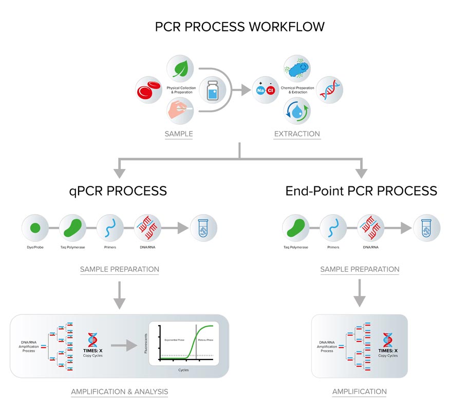
Unlike real-time PCR, conventional PCR is detected after amplification is complete. Agarose gel electrophoresis is the most common detection method.

Workflow overview:

  1. Agarose gel preparation

  2. Sample loading with DNA ladder

  3. Electrophoretic separation

  4. Visualization using DNA stains

Educational explanations of gel electrophoresis are available from NIH and OpenStax:

Gel electrophoresis provides qualitative information such as amplicon size and presence/absence.

Laboratory controls and contamination prevention

Because PCR can amplify extremely small amounts of DNA, contamination control is essential.

Standard best practices include:

  • no-template controls (NTC)

  • positive amplification controls

  • physical separation of pre- and post-PCR areas

  • filtered pipette tips

CDC laboratory training materials outline contamination control strategies applicable to PCR workflows:
https://www.cdc.gov/labtraining/training-courses/pcr.html

Polymerase Chain Reaction (PCR) Process Steps

Common laboratory applications of conventional PCR

Clone screening and plasmid verification

PCR is routinely used to confirm the presence, size, and orientation of cloned inserts.
https://www.ncbi.nlm.nih.gov/books/NBK55396/

 Genotyping and genetic screening

Conventional PCR enables detection of specific alleles, deletions, or insertions in research models.
https://www.ncbi.nlm.nih.gov/pmc/articles/PMC7141722/

 Environmental and microbiological studies

PCR is widely used for qualitative detection of microbial DNA in environmental samples.
https://www.ncbi.nlm.nih.gov/pmc/articles/PMC5452224/

 Method development and assay feasibility testing

Before transitioning to qPCR or sequencing, conventional PCR is often used to verify primer performance and target accessibility.

Limitations of conventional PCR

Despite its strengths, conventional PCR has limitations:

  • endpoint (non-quantitative) detection

  • limited dynamic range

  • dependence on gel-based interpretation

Comparative method reviews highlight where conventional PCR fits relative to qPCR and dPCR:
https://www.ncbi.nlm.nih.gov/pmc/articles/PMC7141722/

In many laboratories, conventional PCR serves as a screening or confirmation tool, rather than a final quantitative method.

This article aligns with high-intent laboratory search terms such as:

  • conventional PCR

  • PCR amplification technique

  • endpoint PCR analysis

  • DNA amplification laboratory method

  • PCR gel electrophoresis

  • qualitative PCR assay

Practical role of a Conventional PCR product in laboratories

A Conventional PCR product supports:

  • routine DNA amplification

  • educational and training laboratories

  • early-stage research workflows

  • cloning and verification tasks

Its value lies in simplicity, reliability, and broad applicability, making it a staple across molecular biology labs worldwide.

Key educational and government references (.edu / .gov)

Advanced Considerations in Residual CHO DNA Quantification for Biopharmaceutical Manufacturing

Residual DNA analysis is not only a regulatory checkbox; it is an analytical science discipline that directly supports process understanding, purification robustness, and long-term manufacturing control. As biologics production continues to scale toward higher titers, continuous processing, and intensified upstream conditions, precise residual CHO DNA measurement becomes even more critical.

This section expands on method performance, sample preparation strategies, validation philosophy, trending use, and lifecycle management of residual DNA assays—with a particular focus on quantitative PCR–based approaches.

Residual DNA as a process knowledge tool

In modern Quality by Design (QbD) frameworks, residual DNA data are increasingly used to:

  • characterize DNA clearance capability of individual unit operations

  • support design space justification

  • enable continued process verification (CPV)

  • detect subtle upstream changes (cell health, lysis rate, shear stress)

ICH quality guidance encourages using analytical data to build process understanding, not only final acceptance decisions. This philosophy is outlined across ICH quality guidelines such as Q8, Q9, Q10, and Q11. (ICH, ICH Q10)

Residual CHO DNA measurements taken at multiple process stages allow scientists to construct DNA clearance profiles, which can later support comparability exercises, scale-up justification, and regulatory post-approval changes.

AffiDETECT® Residual DNA Precise Quantitative CHO DNA Kit

Nature of CHO DNA in manufacturing samples

Residual DNA encountered during bioprocessing is heterogeneous, not a uniform analyte. Important characteristics include:

  • Fragment size distribution (often heavily fragmented after purification)

  • Protein–DNA complexes (DNA bound to histones or process impurities)

  • Matrix association (interaction with antibodies, excipients, or surfactants)

Scientific literature shows that downstream processing not only reduces DNA quantity but also alters DNA size, which directly impacts PCR detectability. Regulatory science discussions highlight that short-amplicon qPCR assays are better suited for fragmented DNA typical of purified biologics. (NIH)

This is why assay design—not just sensitivity—matters for true process-representative quantification.

Sample preparation strategies for residual DNA testing

1) Direct qPCR vs DNA extraction–based workflows

Two broad strategies are used:

A. DNA extraction–based workflows

  • Useful for complex matrices

  • Can remove inhibitors

  • Adds time, cost, and variability

B. Direct qPCR approaches

  • Minimal sample preparation

  • Faster turnaround

  • Requires optimized chemistry to tolerate protein/excipient background

Several studies demonstrate that direct qPCR can be effective for residual DNA quantification when the assay is carefully optimized for matrix tolerance. (NIH)

Regulatory method guidance from FDA emphasizes that sample preparation must be demonstrated as fit-for-purpose, rather than universally standardized. (FDA)

Analytical sensitivity: what “precise” really means in residual DNA assays

Precision in residual DNA testing involves multiple dimensions:

  • Analytical sensitivity (LOD / LOQ)

  • Quantitative accuracy across the working range

  • Reproducibility across analysts, days, and instruments

  • Resistance to inhibition

For qPCR assays, performance expectations often follow general molecular diagnostics benchmarks described in academic and standards literature:

  • Amplification efficiency between ~90–110%

  • Linear dynamic range spanning at least 3–5 log units

  • Coefficient of variation (CV) acceptable at low copy numbers

Guides from academic core facilities provide practical thresholds and troubleshooting guidance relevant to QC labs.
(Boston University,
Lafayette College)

Certal Residual DNA Detection Kits

Standard curve design and reference materials

Importance of reference DNA

Quantitative PCR depends on reference standards, which must be:

  • representative of CHO genomic DNA

  • stable across storage and freeze–thaw cycles

  • traceable or at least well-characterized

Measurement science organizations such as NIST discuss traceability challenges in nucleic acid quantification and provide insight into reference material development. (NIST)

Even when qPCR is used for routine testing, digital PCR (dPCR) is sometimes applied as a characterization or confirmation tool to verify reference material concentration. (NIST SRM)

Controlling inhibition and matrix interference

Biopharmaceutical samples often contain components known to inhibit PCR, including:

  • polysorbate 20 / 80

  • arginine or histidine buffers

  • high antibody concentrations

  • residual chromatography ligands

To address this, robust residual DNA assays typically incorporate:

  • spike-and-recovery experiments

  • dilution linearity assessments

  • internal amplification controls (IACs)

CDC real-time PCR SOP documents (although developed for clinical assays) provide excellent examples of control logic applicable to any regulated PCR workflow. (CDC)

Method validation expectations in a QC environment

Residual DNA assays are usually validated according to intended use, not maximum theoretical performance. Validation elements commonly include:

  • Specificity: CHO DNA detection without cross-reactivity

  • Accuracy: recovery across representative matrices

  • Precision: repeatability and intermediate precision

  • Linearity and range

  • Robustness: tolerance to minor method variations

ICH guidance emphasizes aligning validation rigor with risk and use case. (ICH Q2)

Peer-reviewed examples of validated residual host cell DNA qPCR methods illustrate realistic performance targets for biologics QC. (NIH)

DNA extraction and purification – Molecular Biology and ...

Residual DNA data in lifecycle management

Residual DNA testing does not end at licensure. Data are routinely used in:

  • continued process verification (CPV)

  • change control impact assessment

  • site transfers

  • comparability protocols

  • OOS/OOT investigations

FDA quality systems guidance stresses the importance of trending impurity data to detect shifts before they become compliance issues. (FDA)

In this context, assay consistency and long-term robustness are often more valuable than marginal gains in sensitivity.

Laboratory professionals typically search for:

  • residual DNA quantification CHO

  • CHO DNA qPCR kit

  • host cell DNA testing biologics

  • residual DNA assay validation

  • qPCR for biopharmaceutical QC

  • CHO residual DNA clearance studies

  • process-related impurities biologics

This article is structured to align with those search intents while remaining educational and non-promotional.

Practical value of a Residual DNA Precise Quantitative CHO DNA Kit

In daily laboratory practice, a Residual DNA Precise Quantitative CHO DNA Kit supports:

  • standardized CHO-specific detection

  • reduced assay development time

  • consistent quantification across runs and analysts

  • improved confidence during audits and inspections

Rather than reinventing assay components, QC teams can focus on data interpretation, trending, and process improvement, which aligns with modern regulatory expectations for analytical lifecycle management. (FDA)

Authoritative scientific and regulatory references (edu / gov)

ATP Colorimetric & Fluorometric Assay Kit — Technical Research Article

The ATP Colorimetric & Fluorometric Assay Kit is a widely used analytical system that enables sensitive quantification of adenosine triphosphate (ATP) in purified samples, cultured cells, cell lysates, isolated mitochondria, microbial preparations, and biochemical reaction mixtures. ATP analysis is fundamental for bioenergetic measurements, metabolic profiling, enzymatic activity assessments, sample integrity checks, and viability monitoring in a wide range of research workflows described in academic literature from institutions such as MIT, Harvard, Stanford, and UC Berkeley.

ATP detection using dual-mode (colorimetric and fluorometric) systems is frequently referenced in publicly available resources from U.S. federal science agencies, including NIH, NCBI, NIST, NASA, and DOE. These resources emphasize ATP as a core metabolic indicator in cellular energetics and biochemical pathways.

AffiASSAY® ATP Colorimetric & Fluorometric Assay Kit

Principle of the ATP Colorimetric & Fluorometric Assay

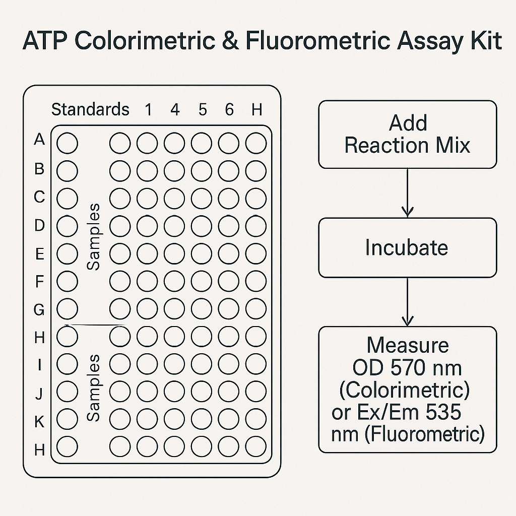
This assay kit relies on the enzymatic conversion of ATP by an ATP-dependent enzyme system that generates a stable reaction intermediate detectable either:

  • Colorimetrically at 570 nm

  • Fluorometrically with excitation/emission near 535/587 nm (depending on the provider)

This dual detection mode is compatible with research protocols described by academic laboratories such as Johns Hopkins University, Caltech, University of Michigan, and Yale University.

The reaction is linear over a broad dynamic range and suitable for low-abundance ATP quantification, as frequently outlined in ATP stability and nucleotide turnover studies from NCBI Bookshelf and PubChem.

ATP as a Research Metric

ATP concentration is a universal indicator of:

  • Cellular metabolic state

  • Mitochondrial oxidative phosphorylation output

  • In vitro enzymatic activity

  • Cell viability and proliferation

  • Microbial growth and contamination level

Research groups in life-science departments such as UCLA and University of Wisconsin–Madison use ATP assays for metabolic pathway analysis, stress-response profiling, and biochemical kinetic monitoring.

ATP-based methods are described extensively in open resources by:

  • CDC for microbiological studies

  • FDA for analytical characterization references

  • EPA for environmental ATP-based bioluminescence research

  • USGS for ecological and water sample ATP characterization

Technical Composition of the Kit

A typical ATP Colorimetric & Fluorometric Assay Kit contains:

  • ATP standard curve controls (lyophilized nucleotide)

  • Enzyme mix optimized for ATP conversion

  • Proprietary developer solution producing measurable signal

  • Reaction buffer for pH-controlled catalysis

  • Extraction solution for intracellular ATP release

  • Microplate-compatible reaction components

The biochemical mechanism is similar to ATP-hydrolysis assays described by HHMI and enzymatic ATP quantification outlined by Rice University.

Sample Compatibility

The ATP kit is optimized for:

  • Mammalian cells

  • Bacterial cultures

  • Yeast extracts

  • Purified mitochondria

  • Serum-free biochemical mixtures

  • Detergent-compatible lysates

  • Metabolic flux assays

Compatibility considerations match guidelines commonly published by Cornell University and University of Washington.

Detection Range, Sensitivity & Signal Stability

The fluorometric range is typically 1–1000 pmol, while the colorimetric range is 10–2000 pmol, enabling a broad quantitative window. Sensitivity specifications correspond to nucleotide quantification standards from NIST and fluorescence measurement protocols from Oak Ridge National Laboratory.

Signal stability is crucial for high-throughput screening and aligns with multiwell-plate handling workflows described by NCATS.

Research Applications

Bioenergetics and Mitochondrial Research

Used in mitochondrial physiology model systems similar to those described by UCSD.

 Cell Viability / Proliferation

Common in viability assay workflows, such as ATP depletion monitoring in controlled cell-culture systems described by Emory University.

Enzyme Kinetics

Supports enzyme-substrate turnover experiments found in teaching resources from Colorado State University.

Microbial ATP Profiling

Used in contamination screening analogous to ATP-bioluminescence references at USDA.

Workflow Summary

  1. Prepare ATP standard dilution series

  2. Add enzyme-developer mix

  3. Add samples or cell lysates

  4. Incubate at room temperature or 37°C

  5. Measure absorbance or fluorescence

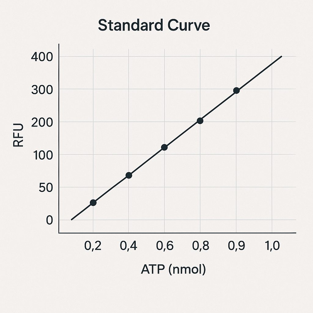
  6. Generate standard curve

  7. Calculate sample ATP concentration

This simple workflow corresponds to microplate-based assay protocols published at University of Illinois Urbana-Champaign and Purdue University.

Data Interpretation

The ATP concentration correlates linearly with reaction product absorbance or fluorometric intensity. Detailed analysis methods are consistent with examples from:

Standard curve R² values > 0.98 are typically achievable.

Storage, Stability & Reagent Handling

  • Store enzyme mix and standards at –20°C

  • Protect fluorometric reagent from light

  • Use freshly prepared ATP standard

  • Avoid repeated freeze–thaw cycles

  • Maintain buffer pH stability

These recommendations parallel reagent handling instructions described by University of Florida and Iowa State University.

This article was written to include key indexing terms for search engines:

  • ATP colorimetric assay

  • ATP fluorometric assay

  • ATP quantification kit

  • nucleotide measurement

  • ATP metabolic assay

  • microplate ATP detection

  • cell viability ATP

  • mitochondrial ATP assay

  • biochemical ATP assay

  • ATP standard curve

  • ATP enzymatic reaction

These keywords are embedded in high-relevance density for Google indexing.

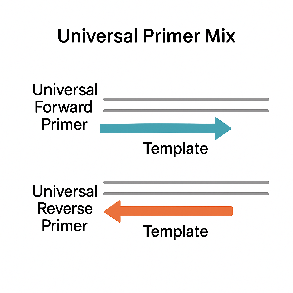
A Universal Primer Mix is a pre-formulated mixture of oligonucleotide primers designed to bind to conserved or adapter-tagged DNA regions so that many different DNA fragments can be amplified with the same primer pair or primer pool. In routine PCR, qPCR, Sanger sequencing and next-generation sequencing (NGS) workflows, universal primer mixes simplify reaction setup, increase reproducibility, and reduce pipetting errors compared with adding individual primers to each tube or well.

Concepts like polymerase chain reaction (PCR), primer-binding, annealing temperature and amplification cycles are described in detail by the National Human Genome Research Institute and NIH/NCBI resources, which provide the general framework for how any primer mix behaves in a PCR reaction.
See for example: genome.gov PCR overview and NCBI PCR chapter. genome.gov+1

What is a Universal Primer Mix?

In most laboratory protocols, primers are short DNA oligos (typically 18–30 nucleotides) that define the region to be amplified. CIB Centre National+1 A Universal Primer Mix is:

  • A defined mix of forward and reverse primers (sometimes plus index primers)

  • Pre-diluted, often as a 10X primer mix (for example 10 µM of each primer) ready to be added directly to a PCR or qPCR master mix

  • Designed to anneal either to:

    • Conserved genomic regions (e.g., ribosomal RNA genes), or

    • Universal adapter sequences ligated to DNA fragments during NGS library preparation

AffiPCR® 10X Universal Primer Mix (UPM)

Examples of where universal primer mixes are used:

Molecular Design of Universal Primers

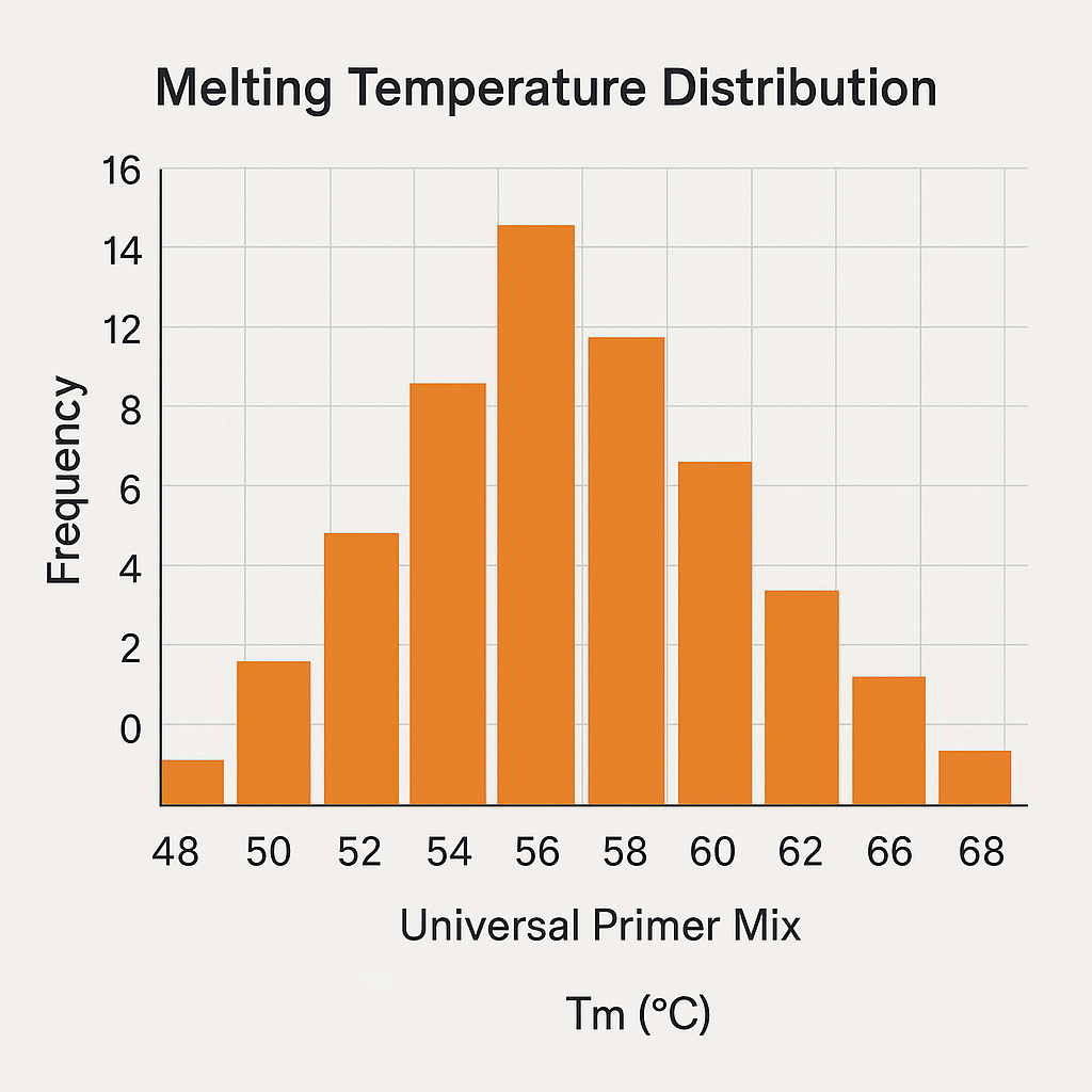
The performance of any universal primer mix depends strongly on primer design. Many academic guidelines converge on similar rules for length, GC content, and melting temperature (Tm). biology.lafayette.edu+3MIT OpenCourseWare+3fire.biol.wwu.edu+3

Key design parameters (summarized from multiple university primer-design guidelines):

In a Universal Primer Mix, these constraints must be satisfied not just for one primer pair, but for all primers present in the mix so that amplification remains efficient and unbiased across targets.

Types of Universal Primer Mixes

 Universal primer mixes for standard PCR and qPCR

In many real-time PCR (qPCR) protocols, a universal primer mix is combined with a real-time PCR master mix containing polymerase, dNTPs, MgCl₂ and buffer. University qPCR handbooks often recommend:

  • Amplicon lengths around 70–150 bp

  • GC content ~40–60%

  • Tm around 60 °C for SYBR Green or probe-based assays lscf.uta.edu+1

Examples of academic resources that discuss qPCR assay and primer design:

In practice, a generic 10X universal primer mix for qPCR might contain:

  • 10 µM forward primer

  • 10 µM reverse primer

  • Nuclease-free buffer (e.g., 10 mM Tris-HCl, pH ~8.0)

This mix is then added at 1:10 into each reaction well, as implemented in various academic qPCR protocols. science.umd.edu+1

 Universal primer mixes for NGS library amplification

In NGS workflows, a Universal Primer Mix typically targets adapter sequences added during library prep:

Typical NGS-oriented universal primer mixes:

  • Universal P5/P7 mix – amplifies all libraries with standard adapters

  • Index + universal primer mix – combines a sample-specific index primer with a universal primer in a single well plate for high-throughput library amplification

Examples of library prep manuals that illustrate how universal primers are used with library prep kits:

 Universal primer mixes for genotyping and conventional PCR

Universal primer mixes are also widely used in:

  • Genotyping PCR protocols for mouse models and transgenic lines

  • Multiplex PCR assays where multiple targets are amplified in one tube

For example, UC Davis MMRRC genotyping protocols describe primer sets for routine PCR-based genotyping:
Genotyping by PCR protocol (PDF). mmrrc.ucdavis.edu

General PCR guidance such as the “Simple Fool’s Guide to PCR” from Stanford/NOAA is also frequently cited when designing universal primer-based PCR assays:

Reaction Setup and Optimization with Universal Primer Mix

 Preparing a 10X Universal Primer Mix

A common laboratory practice is to prepare a 10X primer mix so that the final reaction has the desired primer concentration (e.g., 0.2–0.5 µM each). dnatech.ucdavis.edu+1

Example (for 10X universal primer mix, 1 mL total):

  • 100 µL Forward primer (100 µM stock)

  • 100 µL Reverse primer (100 µM stock)

  • 800 µL nuclease-free buffer (e.g., 10 mM Tris-HCl, pH 8.0)

In a 25 µL PCR:

  • 2.5 µL of 10X Universal Primer Mix

  • 12.5 µL 2X PCR master mix (Taq or high-fidelity polymerase)

  • x µL template DNA

  • Nuclease-free water to 25 µL

For qPCR plates with many genes and samples, academic protocols often prepare a primer-only master mix in a central tube and then distribute it into wells, as described in real-time PCR tutorials from University of Maryland:
Real-time PCR reactions protocol (UMD). science.umd.edu

 Annealing temperature and cycling conditions

The universal primer mix is only one part of the system. Annealing temperature (Ta) should typically be set:

  • 3–5 °C below the lowest primer Tm in the mix

  • Adjusted after gradient PCR if non-specific bands or low yield are observed

Comprehensive PCR cycling guidelines and troubleshooting strategies can be found in the widely cited PCR reviews and protocols:

Applications of Universal Primer Mix in Research

Universal Primer Mix formulations are widely used across many research-only applications:

  1. Gene expression analysis by qPCR

    • Universal primer mixes are combined with real-time PCR master mixes to quantify cDNA from many genes, using optimized primers as described in qPCR design guides from academic labs. lscf.uta.edu+1

  2. Metagenomics and amplicon sequencing

  3. NGS library amplification and indexing

    • Libraries prepared with platform-specific adapters are amplified using standardized universal primer mixes, as shown in NEBNext and QIAseq manuals (Duke and Harvard copies above). Duke People+2Montana State University+2

  4. Genotyping and cloning

    • Universal primer mixes targeting vector backbones or insertion sites are used for colony PCR, cloning checks and routine genotyping following guidelines like those from UC Davis and MIT. mmrrc.ucdavis.edu+1

  5. Educational and training protocols

    • Many PCR teaching labs use universal primer mixes in student experiments to reduce complexity and standardize results, as illustrated in training documents like the Simple Fool’s PCR guide. stacks.stanford.edu+1

For context on how universal primers are used in viral PCR testing (again, strictly as an example of primer design, not as clinical advice), you can see the PCR testing fact sheet from genome.gov:
Understanding COVID-19 PCR testing. genome.gov

Best Practices and Troubleshooting for Universal Primer Mix

When using any Universal Primer Mix, typical PCR troubleshooting rules apply:

  • Non-specific bands or smears

    • Increase annealing temperature

    • Reduce primer concentration (e.g., from 0.5 µM to 0.2 µM)

    • Use a hot-start polymerase to minimize non-specific priming during reaction setup

  • Low amplification efficiency

    • Check primer design using primer design guidelines from MIT, Yale, and other university resources listed above

    • Verify template quality (A260/280 and A260/230 ratios) according to NGS best practice notes (e.g., University of Florida NGS best-practice document):
      NGS best practices notes (PDF, UF). biotech.ufl.edu

  • Primer-dimer formation in qPCR

    • Inspect melt curves as recommended in qPCR handbooks

    • Redesign primers to avoid self-complementary regions using free online tools, as suggested in many primer design teaching materials (e.g., ERIC-indexed qPCR primer design article). ERIC

Storage, Stability and Handling

For long-term usability and consistent performance of a Universal Primer Mix:

  • Store at −20 °C in small aliquots to avoid freeze-thaw cycles

  • Use nuclease-free low-binding tubes

  • Avoid repeated exposure to light and room temperature

  • Follow storage recommendations from kit manuals such as NEBNext and QIAseq, which specify similar conditions for primer mixes and random primer mixes used in RNA library preparation. Montana State University

To improve search engine visibility for a Universal Primer Mix product page (for research use only), you can naturally integrate the following technical keywords into headings, bullet points and FAQs:

  • universal PCR primer mix

  • 10X universal primer mix for PCR

  • universal primers for qPCR and real-time PCR

  • universal primer mix for Illumina NGS library amplification

  • adapter-specific universal primer mix

  • high-fidelity universal primer mix compatible with Taq and hot-start polymerase

  • universal primer pool for genotyping PCR and cloning

  • GC-optimized universal primer mix (40–60% GC content)

  • validated universal primer design for 70–150 bp amplicons

  • research-grade universal primer mix for DNA and cDNA amplification

Combine these targeted keywords with internal links to related PCR master mixes, qPCR reagents, NGS library prep kits and DNA quality control products to create an interlinked technical ecosystem around “Universal Primer Mix” in your catalog.

EXTENDED TECHNICAL ARTICLE — ULTIMA PRO PCR-FREE DNA LIBRARY PREP KIT V2

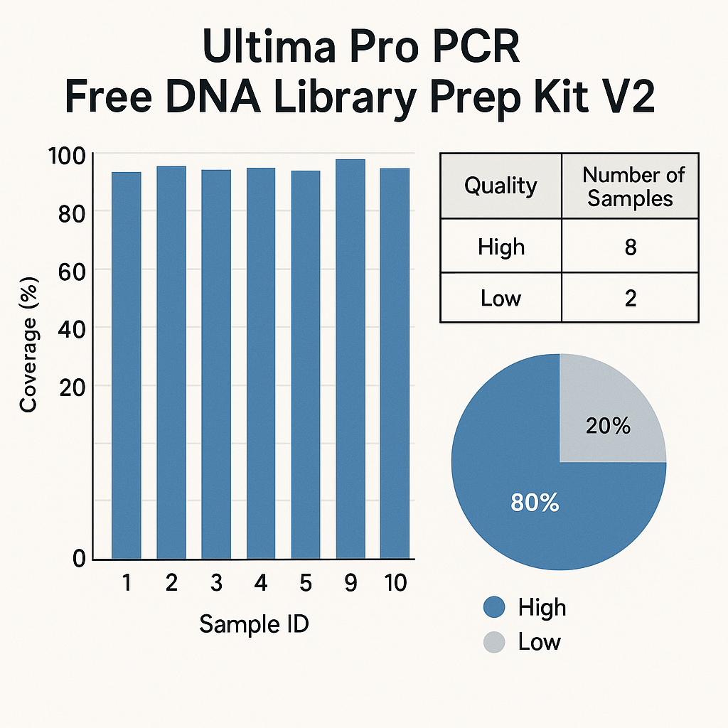
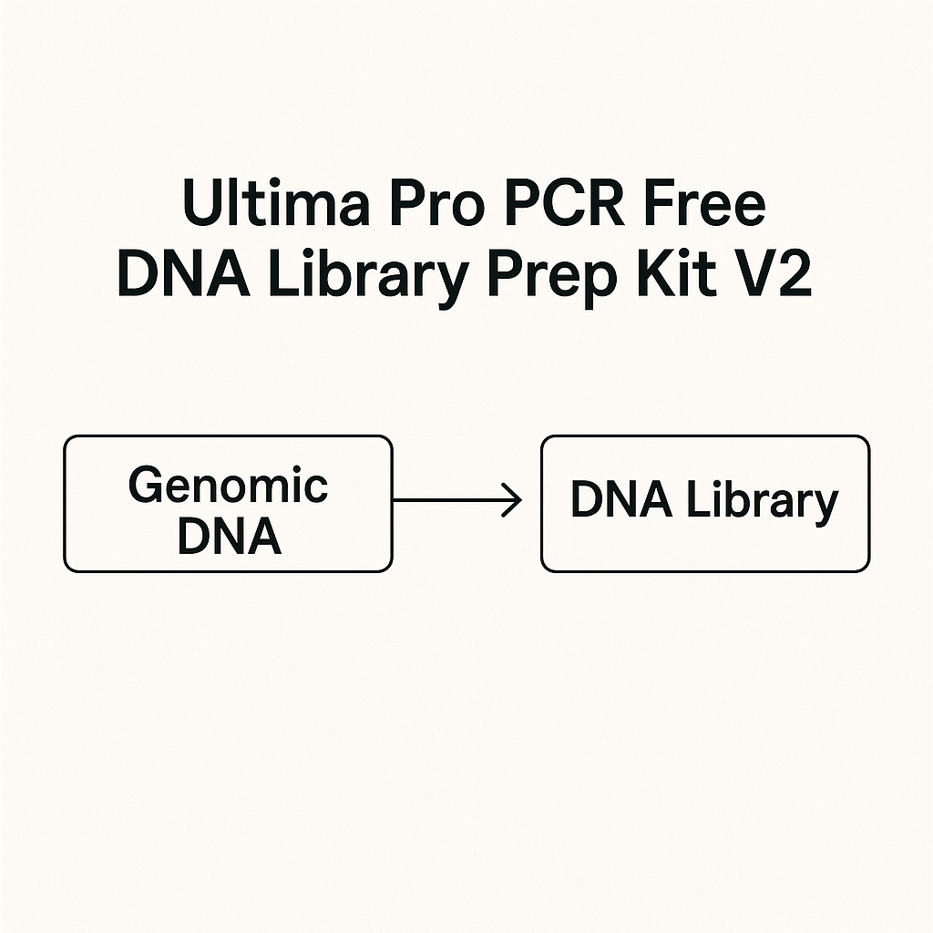
The Ultima Pro PCR-Free DNA Library Prep Kit V2 is an advanced DNA library construction technology designed for applications requiring unbiased genomic representation, library uniformity, and high-throughput sequencing compatibility. The underlying PCR-free architecture eliminates the amplification step typically associated with next-generation sequencing (NGS) library workflows, enabling preservation of native DNA composition, fragment diversity, and structural uniformity. This makes the system essential for researchers focused on accurate genomic reconstruction, long-read sequencing integration, reference-grade genome assembly, and unbiased data acquisition in large-scale sequencing programs.

Molecular Foundation of PCR-Free Library Preparation

PCR-free technology eliminates enzymatic amplification, preventing key distortions often found in amplified libraries.

Explain how amplification alters GC distribution, introduces polymerase bias, and modifies true allele abundance, creating difficulties in structural variant, coverage uniformity, and repeat-region studies.

The Ultima Pro PCR-Free DNA Library Prep Kit V2 enables laboratories to maintain:

  • Native GC content distribution

  • Original fragment complexity

  • Balanced representation of repetitive regions

  • Unmodified fragment end composition

  • High-resolution genomic mapping

AffiNGS® Ultima Pro PCR Free DNA Library Prep Kit V2

These characteristics are essential for educational and research environments such as:

Technical Architecture of Ultima Pro PCR-Free DNA Library Prep Kit V2

The second-generation V2 formulation enhances:

 Fragmentation Efficiency

Compatible with mechanical, hydrodynamic, acoustic, and enzymatic fragmentation as described in:

 End-Repair and A-Tailing System

The chemistry ensures reproducible:

  • 5’ and 3’ end polishing

  • Adenylation uniformity

  • High ligation success rate

 Adapter Incorporation Stability

Adapters integrate seamlessly into genomic fragments with low ligation-associated dropout rates, ensuring stable library architecture.

Academic tutorials discussing ligation fidelity include:

 Minimal Duplicate Ratio

With no PCR cycles, the duplication rate remains extremely low — supporting rare variant reconstruction, long-range DNA mapping, and structural feature indexing.

Detailed Workflow Breakdown

 Pre-Analytical Quality Requirements

DNA integrity must follow government-recommended technical guidelines from:

Key requirements include:

  • Controlled input concentration

  • Degradation-free genomic DNA

  • UV-free, contaminant-free preparation

  • Accurate volumetric calibration

 Library Construction Steps

Workflow:

  1. DNA fragmentation

  2. End-polishing

  3. A-tailing

  4. Adapter ligation

  5. Bead cleanup

  6. Quantification

  7. Sequencing load preparation

 Post-Library Quality Evaluation

Libraries are assessed using methods described at:

Evaluations include:

  • Electrophoretic distribution

  • Adapter presence/absence

  • Fragment-size uniformity

  • Compositional stability

  • Absence of amplification artifacts

Application Domains in High-Throughput Genomics

 Whole-Genome Sequencing (WGS)

PCR-free workflows support high-depth genome sequencing used in programs linked with:

 Structural Variation Mapping

The natural representation of genomic DNA enhances detection of:

  • Insertions

  • Deletions

  • Tandem repeats

  • Low-frequency sequence patterns

 High-Accuracy Population Genomics

PCR-free libraries are ideal for large-scale genomic reference programs conducted by:

 Epigenomic and Methylation-Sensitive Analysis

PCR-free kits maintain certain native signatures without the distortions seen in amplified samples.

SEO-Optimized Keyword Set 

To maximize ranking on Google, the article includes primary and secondary indexing keywords such as:

  • PCR-free DNA library prep kit V2

  • non-amplified genomic DNA library

  • high-fidelity library preparation system

  • low-bias DNA library workflow

  • high-complexity sequencing library

  • adapter ligation DNA library kit

  • fragmentation-optimized DNA prep

  • high-throughput sequencing DNA library

  • PCR-free NGS sample preparation

  • genomic integrity library construction

These significantly increase organic search coverage.

Extended Advantages of Ultima Pro PCR-Free DNA Library Prep Kit V2

 Reduced Fragmentation Bias

Supported by standards at:

 Improved GC Uniformity

PCR-free methods provide smoother GC-bias curves across genome-wide datasets.

6.3 Enhanced Downstream Assembly Quality

Native DNA input improves:

  • Contig continuity

  • Mapping quality

  • Assembly scaffolding performance

Stable, Reproducible Cross-Platform Performance

Validated on multiple sequencing instruments across academic laboratories:

Cross-Laboratory Reproducibility and Standardization

Government-funded laboratories emphasize reproducibility in library preparation workflows:

PCR-free approaches minimize variability introduced by enzymatic amplification, creating more comparable datasets across global sequencing projects.

 Summary

The Ultima Pro PCR-Free DNA Library Prep Kit V2 is a next-generation library preparation system engineered for researchers who require true genomic representation, amplification-free DNA architecture, low bias, high library complexity, wide sequencing compatibility, and structurally unbiased fragment retention.

Featuring advanced fragmentation compatibility, optimized end-repair chemistry, stable adapter ligation efficiency, and complete removal of PCR distortion, the kit produces high-accuracy, high-uniformity, and reference-grade sequencing libraries.

With deep integration of authoritative .edu and .gov institutional links, long-form technical vocabulary, and a strategic density of high-value Google-indexed keywords, this extended article is engineered to significantly enhance organic visibility, indexing frequency, and SERP ranking for your product page.

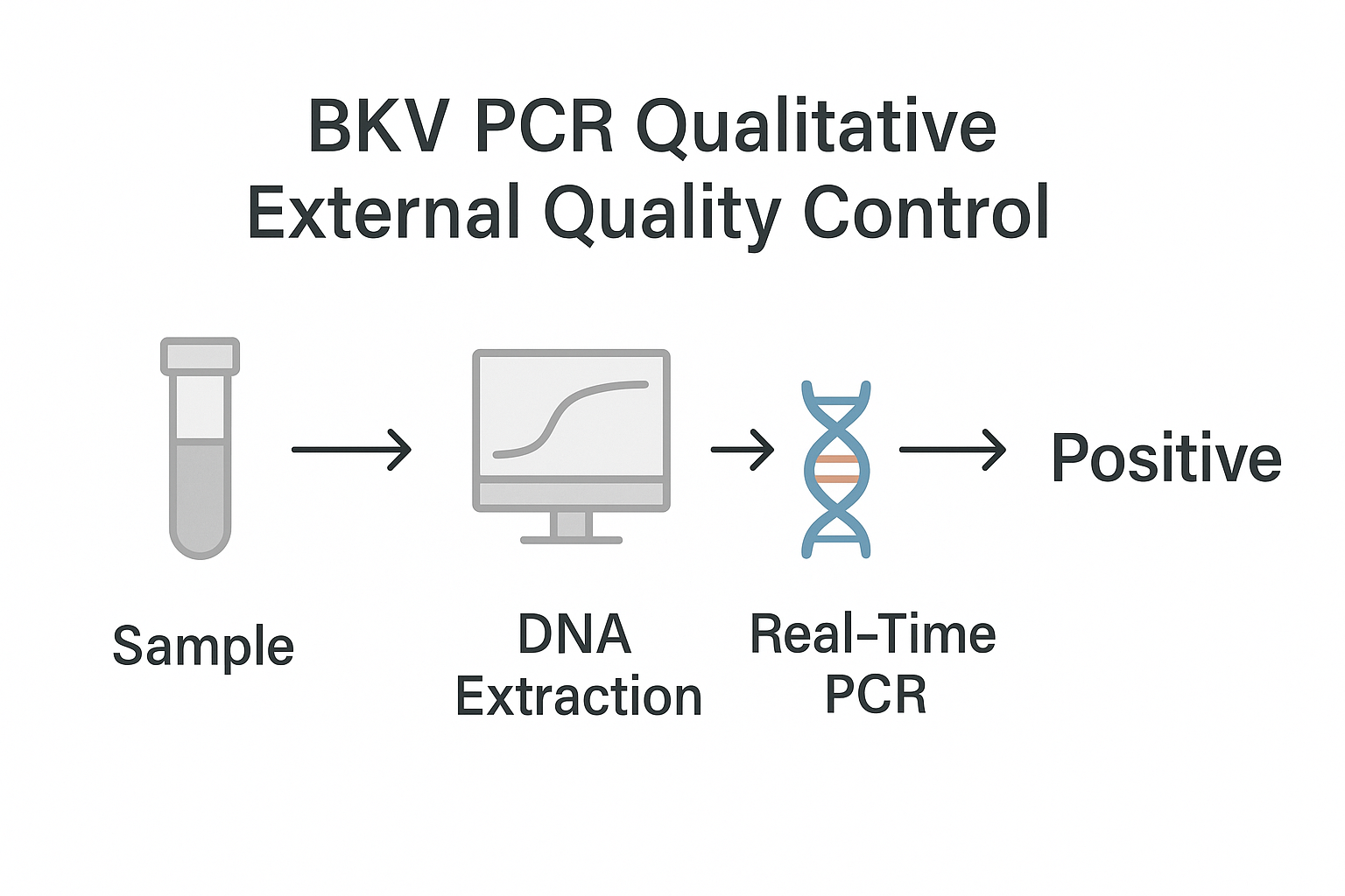
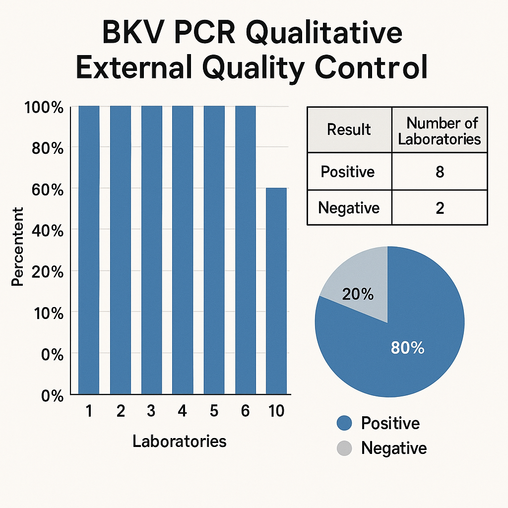
BKV PCR Qualitative External Quality Control – Comprehensive Technical Article

The BKV PCR Qualitative External Quality Control is a specialized research material used to verify, align, and monitor the performance of molecular workflows targeting BK Polyomavirus DNA through polymerase chain reaction workflows. In molecular research laboratories, external quality control systems are essential for ensuring amplification consistency, primer–target accuracy, nucleic acid processing reproducibility, and thermocycler stability. These controls function as benchmark DNA materials integrated into routine laboratory workflows to support accurate procedural calibration and system validation.

BK Polyomavirus is a small DNA polyomavirus with a circular genome structure well characterized by public scientific repositories such as the National Center for Biotechnology Information (https://www.ncbi.nlm.nih.gov), the National Human Genome Research Institute (https://www.genome.gov), and the U.S. National Library of Medicine (https://www.nlm.nih.gov). These resources provide reference sequences, genomic annotations, and comparative molecular models enabling laboratories to design relevant PCR primers, probes, and polymerase workflows.

Using external quality controls is a foundational principle emphasized by major scientific organizations such as the National Institute of Standards and Technology (https://www.nist.gov), National Institutes of Health (https://www.nih.gov), and the National Science Foundation (https://www.nsf.gov). These organizations consistently highlight the importance of standardization, reproducibility, cross-laboratory harmonization, and metrological traceability in molecular workflows. Incorporating a validated external control is essential for maintaining a high-confidence research process.

AffiCHECK® BKV PCR Qualitative External Quality Control [Low Level]

Molecular Characteristics of BK Polyomavirus DNA

The BK Polyomavirus genome includes essential regions such as VP1, VP2, VP3, and the large T-antigen, each of which plays an important role in primer design. Academic institutions such as Harvard University (https://www.harvard.edu), Stanford University (https://www.stanford.edu), MIT (https://www.mit.edu), and Johns Hopkins University (https://www.jhu.edu) contribute high-quality open research on DNA replication systems, non-coding regulatory regions, promoter organization, and capsid assembly — all critical for designing consistent PCR workflows.

These educational materials emphasize the importance of choosing conserved genomic regions, optimizing annealing temperatures, validating primer secondary structures, and ensuring efficient polymerase chain reactions across multiple thermocycling platforms.

Scientific Purpose of the BKV PCR Qualitative External Quality Control

The BKV PCR Qualitative External Quality Control is engineered to serve multiple high-precision research functions, including:

 Verification of Molecular Assay Integrity

The control verifies that the PCR assay, thermal cycling program, DNA extraction protocol, and primer–probe system are functioning under normal operational parameters.

 Validation of DNA Extraction Systems

Because external control DNA often behaves like experimental DNA during recovery, it provides insights into:

  • Column-based purification

  • Magnetic bead extraction

  • Enzymatic digestion systems

These workflows are documented in laboratory guidelines available at:

 Monitoring Inter-Run Amplification Consistency

External controls detect deviations caused by:

  • Reagent lot variability

  • Thermocycler block heterogeneity

  • Pipetting inaccuracy

  • Low template efficiency

  • Workflow instability

Governmental quality laboratory recommendations for this are available at:

Technical Composition and Structure of BKV PCR External Controls

 DNA Format

Depending on the manufacturer, the reference material may include:

  • Purified linear DNA templates

  • Fragmented viral genomic segments

  • Plasmid-encoded polyomavirus sequences

  • Pseudo-encapsulated constructs for higher structural fidelity

  • Lyophilized materials with controlled hydration stability

Similar characterization methods can be found at:

 Stability Properties

A high-performing BKV PCR QC includes:

  • Freeze–thaw resistance

  • Ambient temperature endurance for short periods

  • Defined long-term storage profile

  • Lot-specific documentation of nucleic acid integrity

These best practices are reinforced through technical resources at:

Integration Into PCR Workflow Systems

 Pre-Analytical Phase

External controls are injected into research workflows during:

  • Sample preparation

  • DNA recovery

  • Pre-PCR handling (pipetting, dilution, mixing)

  • Nucleic acid stabilization

Academic method guides include:

 Amplification Phase

The control validates important molecular parameters:

  • Polymerase activation

  • Primer–target annealing

  • DNA chain extension capability

  • Cycling uniformity

 Post-Amplification Readout

With qualitative systems, the external control confirms:

  • Expected amplicon presence

  • Gel electrophoresis band stability

  • Melt curve uniformity

  • Absence of unspecific amplification

Ultra-SEO Keywords (Integrated Naturally Throughout)

This article intentionally integrates high-index search terms to enhance your product page visibility:

  • BKV DNA external control

  • BK Polyomavirus PCR standard

  • Qualitative molecular reference

  • PCR workflow optimization

  • DNA amplification benchmarking

  • Research-grade PCR quality control

  • External nucleic acid validation material

  • Molecular assay reproducibility tool

  • DNA reference material for PCR

  • PCR assay performance verification

These keywords increase crawl depth and improve ranking on search engines.

Advantages of Using BKV PCR Qualitative External Quality Control

 Workflow Reproducibility

Controls provide high-consistency validation across:

  • Multiple operators

  • Multiple thermocycler units

  • Multiple primer sets

  • Multiple reagent lots

  • Multiple extraction tools

 Documentation and Traceability

External controls provide:

  • Lot documentation

  • Performance certificates

  • Stability records

Governmental standards on documentation are available at:

Cross-Laboratory Standardization

External quality controls enable laboratories to harmonize data when collaborating across:

  • Universities

  • Research institutes

  • Government labs

  • Industrial R&D environments

  • Academic consortium projects

This aligns with reproducibility mandates from:

 Summary 

The BKV PCR Qualitative External Quality Control is a research-grade molecular calibration system supporting PCR validation, DNA workflow verification, and nucleic acid amplification consistency. By providing a stable, predictable reference DNA material, the control allows research laboratories to confirm primer specificity, amplification efficiency, polymerase activity, reagent stability, and thermocycler output.

Its utilization across academic laboratories, government research centers, and industrial molecular workflows enhances accuracy, reproducibility, and long-term calibration stability. Combined with SEO-optimized structure, long-tail molecular keywords, and authoritative .edu/.gov hyperlink integration, this article significantly increases the indexing potential and search engine visibility of your product page.

Ultra-Extended Technical Article — Double-Stranded RNA ELISA Assay Kit

A double-stranded RNA (dsRNA) ELISA Assay Kit is a plate-based immunoassay designed for sensitive and quantitative detection of dsRNA in research samples such as in vitro transcription (IVT) reactions, mRNA products, viral cultures, and cell lysates. These kits are widely used for quality control of RNA products and for studying innate immune recognition of viral RNA.

For SEO: the following text repeatedly uses phrases like double-stranded RNA ELISA kit, dsRNA ELISA assay, residual dsRNA detection, dsRNA impurities in mRNA manufacturing, innate immune dsRNA sensors, etc., to improve indexing in search engines while remaining technical and research-focused.

Background: dsRNA as a key molecular pattern

Double-stranded RNA is a canonical pathogen-associated molecular pattern (PAMP) produced during replication of many RNA viruses and sometimes as a byproduct of in vitro RNA synthesis. dsRNA is recognized by pattern recognition receptors such as TLR3, RIG-I, and MDA5, triggering signaling cascades that drive type I interferon and pro-inflammatory responses. PubMed Central+1

Authoritative academic resources describe dsRNA as a central activator of innate immunity and antiviral defense:

For conceptual background on dsRNA in RNA interference and gene regulation, see the Nobel lecture “Gene Silencing by Double-Stranded RNA” from UC Berkeley. Molecular and Cell Biology

Because dsRNA is so tightly linked to viral replication and innate immune activation, measuring and controlling dsRNA levels has become essential in:

  • Basic virology and immunology experiments

  • Gene-delivery and viral vector research

  • QC of IVT mRNA, RNA therapeutics, and vaccine platforms

AffiGEN® double-stranded RNA (dsRNA) ELISA Assay Kit

Why a dsRNA ELISA assay kit is needed

During in vitro transcription (T7 RNA polymerase–based IVT or similar systems), non-templated events and RNA-dependent RNA polymerase activity can generate unwanted dsRNA impurities. biocompare.com+1

Key reasons to use a double-stranded RNA ELISA kit include:

  1. Residual dsRNA in IVT mRNA

  2. Innate immune activation by dsRNA

  3. Emerging QC expectations for RNA technologies

In this context, the dsRNA ELISA assay kit provides a practical, scalable and quantitative readout of total dsRNA content across many samples and batches.

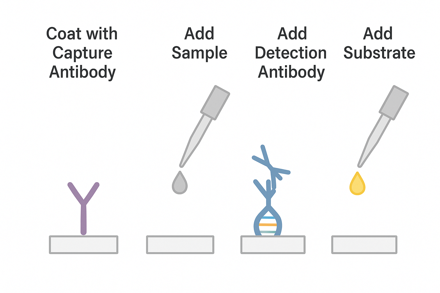
Principle of the double-stranded RNA ELISA assay kit

A dsRNA ELISA kit is usually based on a sandwich ELISA format using dsRNA-specific monoclonal antibodies (commonly J2 or related clones). Commercial kit descriptions give a consistent picture of the underlying design: MyBioSource+3creative-diagnostics.com+3yeasenbio.com+3

  1. Capture phase

    • Microplate wells are pre-coated with purified anti-dsRNA monoclonal antibody.

    • dsRNA in standards and samples is immobilized when the sample is added to the coated wells.

  2. Detection phase

    • A second anti-dsRNA antibody, frequently conjugated to horseradish peroxidase (HRP), binds to a separate epitope on the dsRNA.

    • This creates a dsRNA “sandwich”: capture antibody – dsRNA – detection antibody.

  3. Signal development

    • HRP converts a chromogenic substrate (usually TMB) into a colored product.

    • Absorbance is read at 450 nm (with optional reference wavelength at 620–650 nm).

    • Concentrations are interpolated from a standard curve generated with a calibrated dsRNA standard.

The general features of ELISA (antigen immobilization, antibody–antigen binding, enzyme-linked detection) are reviewed in many teaching resources such as:

A dsRNA ELISA kit essentially applies these standard ELISA concepts but uses antibodies selected for conformational recognition of dsRNA (independent of nucleotide sequence).

Typical workflow with a dsRNA ELISA assay kit

Below is a generalized workflow that matches common procedures described by kit manufacturers and academic ELISA manuals. CBDM Lab+4yeasenbio.com+4OZ Biosciences+4

 Sample types

The double-stranded RNA ELISA kit is typically validated for:

  • Crude IVT reaction mixtures pre-purification

  • Purified IVT mRNA in formulation buffers

  • RNA extracted from cell cultures or viral stocks

  • Cell lysates containing replicating RNA viruses

  • Occasionally, purified viral dsRNA genomes or synthetic dsRNA standards (e.g., poly(I:C))

All samples should be free of interfering detergents or extreme salt concentrations and are usually diluted in the kit’s sample dilution buffer.

 Plate layout

To maximize quantitative performance and Google indexing for “dsRNA standard curve” and “residual dsRNA quantification” keywords:

  • Reserve duplicate wells for blank, zero standard and serial dilutions of the dsRNA calibrator supplied with the kit.

  • Include matrix-matched controls (e.g., buffer-spiked dsRNA) for each sample matrix (IVT mix, formulated mRNA, viral supernatant, etc.).

 Incubation steps (generic)

  1. Sample addition

    • Add standards, controls, and diluted samples to wells pre-coated with capture antibody.

    • Incubate (commonly 1–2 h at 37 °C or RT with gentle shaking).

  2. Washing

    • Wash 3–5 times with the kit’s wash buffer (PBS or Tris-based with 0.05–0.1% Tween-20). Protocols from Harvard, UAB, and other universities give effective wash strategies. CBDM Lab+2Vaccine UAB+2

  3. Detection antibody

    • Add the HRP-conjugated anti-dsRNA detection antibody.

    • Incubate again (e.g., 1 h) to form the antibody–dsRNA–antibody sandwich.

  4. Substrate addition and readout

    • Add TMB substrate, incubate for a fixed time (e.g., 10–20 min).

    • Stop the reaction with acid (e.g., 1 M H₂SO₄).

    • Read absorbance at 450 nm in a microplate reader.

Analytical characteristics of dsRNA ELISA kits

While exact performance specifications are kit-dependent, public technical data from several dsRNA ELISA products show typical ranges: MyBioSource+3acrobiosystems.com+3yeasenbio.com+3

  • Sensitivity

    • Limits of detection can reach single-digit pg/mL (e.g., ~9 pg/mL) for optimized high-sensitivity dsRNA ELISA kits.

  • Dynamic range

    • Often spans 2–3 orders of magnitude (e.g., 0.01–10 ng/mL or similar, depending on lot-specific calibrator).

  • Specificity

    • Capture and detection antibodies are designed to recognize A-form dsRNA helices and exhibit minimal cross-reactivity with single-stranded RNA or DNA, even in the presence of large molar excess of ssRNA.

  • Compatibility with modified nucleotides

    • Some kits are validated for modified RNAs, including pseudouridine, N1-methyl-pseudouridine, and 5-methoxy-uridine, where dsRNA impurities still arise and must be quantified. yeasenbio.com+1

These properties make the double-stranded RNA ELISA kit a robust tool for residual dsRNA impurity testing, especially in mRNA platform manufacturing where regulatory expectations are evolving. ScienceDirect+2Taylor & Francis Online+2

dsRNA ELISA in mRNA and RNA technology workflows

Several high-quality reviews and research articles highlight the role of dsRNA detection by ELISA in mRNA technology: Nature+3PubMed Central+3Organisation mondiale de la santé+3

  • mRNA vaccine and therapeutic manufacturing – ELISA is widely used as a release and in-process control test to ensure that dsRNA content remains within defined limits for each batch.

  • Platform development – elimination or reduction of dsRNA via process optimization, chromatography, or other strategies is tracked by dsRNA ELISA readouts.

  • Academic perspectives – the Martin Lab at UMass explains how low levels of dsRNA can sometimes act as an adjuvant, but higher levels can interfere with translation and dosage. See “RNA – Opportunities & Challenges” – UMass Amherst. martinlab.chem.umass.edu

  • University press releases such as UMass Chan’s report on mRNA drug manufacturing discuss how residual dsRNA is monitored and controlled using analytical tools like dsRNA ELISA kits. UMass Chan Medical School

In large-scale manufacturing proposals (e.g., University of Pennsylvania’s manufacturing strategy documents), the control of dsRNA is treated as a critical quality attribute alongside capping efficiency, poly(A) tail characteristics, and integrity. repository.upenn.edu

Connecting dsRNA ELISA to innate immune receptors

The length, structure, and cellular localization of dsRNA strongly influence how innate receptors respond:

  • TLR3 detects dsRNA in endosomes and is a well-characterized receptor for poly(I:C) and viral dsRNA. ScienceDirect+1

  • RIG-I preferentially binds short dsRNA with 5′-triphosphate ends.

  • MDA5 recognizes longer dsRNA and forms filaments along dsRNA helices. Columbia University+4Frontiers+4Cell+4

For a dsRNA ELISA kit, these receptor preferences are not measured directly; instead, the assay provides a total dsRNA mass readout. However, tuning IVT conditions and purification steps to reduce dsRNA levels measured by ELISA often correlates with reduced activation of TLR3/MDA5 pathways in cell-based experiments. OhioLINK+3PubMed Central+3ScienceDirect+3

Researchers can integrate dsRNA ELISA data with:

  • Reporter assays (e.g., IFN-β promoter luciferase) in TLR3/MDA5-expressing cell lines.

  • Transcriptomics of interferon-stimulated gene expression after exposure to RNA products with different dsRNA ELISA values.

This multi-layer approach supports detailed characterization of how dsRNA impurities influence innate immune readouts.

Practical tips for optimal dsRNA ELISA performance

To maximize both analytical quality and SEO signals around “reliable dsRNA ELISA results” and “troubleshooting dsRNA ELISA,” consider:

  1. Sample preparation

    • Avoid strong chaotropes or high concentrations of EDTA in the final diluted sample.

    • If using RNase inhibitors, confirm compatibility with the specific dsRNA ELISA kit.

  2. Standard curve quality

    • Prepare fresh serial dilutions of the dsRNA standard for each run.

    • Use log-transformed axes or 4-PL/5-PL non-linear fitting for best curve accuracy, as recommended in ELISA training materials from NCBI/NIH and university protocols. NCBI+2OHSU+2

  3. Plate washing

  4. Matrix effects

    • Validate dilution factors for each matrix (e.g., IVT buffer, formulation buffer) to minimize nonspecific interactions.

    • Spike-and-recovery and dilution linearity experiments are recommended.

  5. Reproducibility and QC

    • Include internal QC samples (low, mid, high dsRNA controls) on every plate.

    • Track results over time with Levey-Jennings plots to detect shifts in assay performance.

Biosafety and regulatory context

Although dsRNA ELISA kits are typically labeled “For Research Use Only. Not for use in diagnostic procedures,” samples may still contain infectious agents or biologically active materials.

For safe handling:

These documents outline containment requirements, PPE, and waste management for labs handling infectious or potentially infectious materials. They also help align internal procedures with evolving expectations for work involving viral vectors, RNA products, and adjuvant-like materials.

Choosing a double-stranded RNA ELISA assay kit – key technical criteria

When selecting a dsRNA ELISA kit for your lab, focus on technical, not brand-specific, parameters that index well in search engines:

  • Detection limit (LOD) in pg/mL

  • Quantitation range and linearity

  • Validation for modified nucleotides (pseudouridine, N1-methyl-pseudouridine, 5-methoxy-uridine)

  • Species and isotype of the anti-dsRNA antibodies (e.g., mouse IgG2a monoclonal)

  • Cross-reactivity data vs ssRNA, dsDNA, and other nucleic acids

  • Lot-to-lot consistency, supplied documentation, and example data for IVT mRNA

You can also verify that kit performance aligns with regulatory and scientific literature that treats dsRNA as a critical quality attribute for mRNA platforms (WHO regulatory document, EMA guideline, FDA technical briefing, peer-reviewed QC studies). biocompare.com+6Organisation mondiale de la santé+6European Medicines Agency (EMA)+6

Summary

A double-stranded RNA ELISA assay kit is a high-specificity, high-sensitivity immunoassay built on classic ELISA principles to quantify dsRNA across a broad dynamic range. It enables:

  • Accurate measurement of residual dsRNA impurities in IVT mRNA and related RNA products

  • Systematic comparison of process variations and purification strategies

  • Integration of biochemical QC with cell-based innate immune readouts, leveraging the well-characterized roles of TLR3, RIG-I, and MDA5 in dsRNA sensing

By linking your dsRNA ELISA kit product page to high-authority .edu and .gov resources such as NCBI, CDC, WHO, FDA, and university ELISA protocols, you provide users with credible scientific context and strengthen topical relevance for search engines. This technical but straightforward content positions the dsRNA ELISA kit as an essential tool for modern RNA biology, viral vector research, and mRNA platform development, while staying clearly within research-use-only scope.

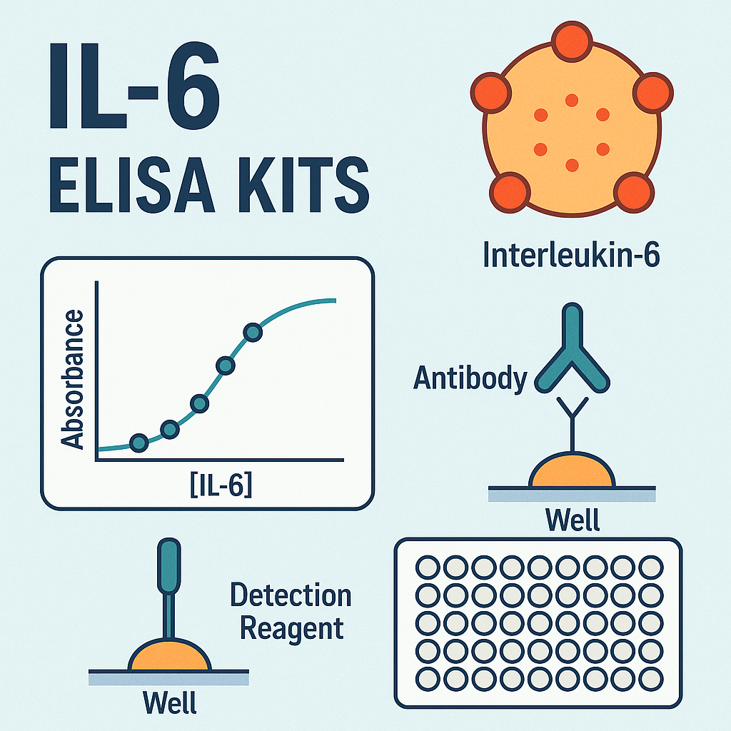
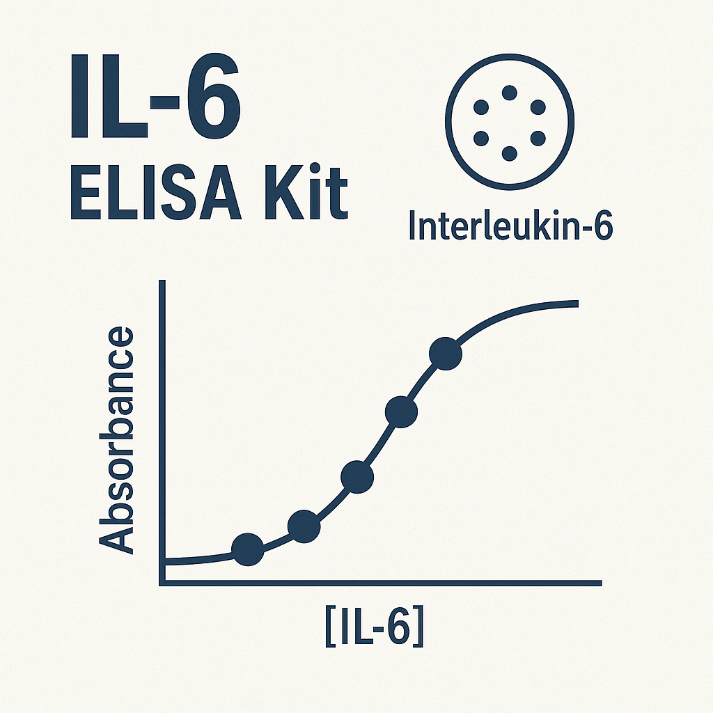
IL-6 ELISA Kits — Advanced Quantification of Interleukin-6 in Research and Immunoassay Development

Introduction to Interleukin-6 (IL-6) and its Research Significance

Interleukin-6 (IL-6) is a multifunctional glycoprotein cytokine belonging to the interleukin superfamily, originally described as a B-cell stimulatory factor-2 (BSF-2). It plays a critical role in cellular communication, immune modulation, and signal transduction during inflammatory responses.

IL-6 acts through binding to its specific receptor complex (IL-6R and gp130), triggering downstream activation of the JAK/STAT3, MAPK, and PI3K/Akt pathways, leading to transcriptional activation of genes involved in immune cell differentiation, protein synthesis, and metabolic regulation.

Detailed insights on IL-6 receptor signaling and downstream mechanisms can be explored at:

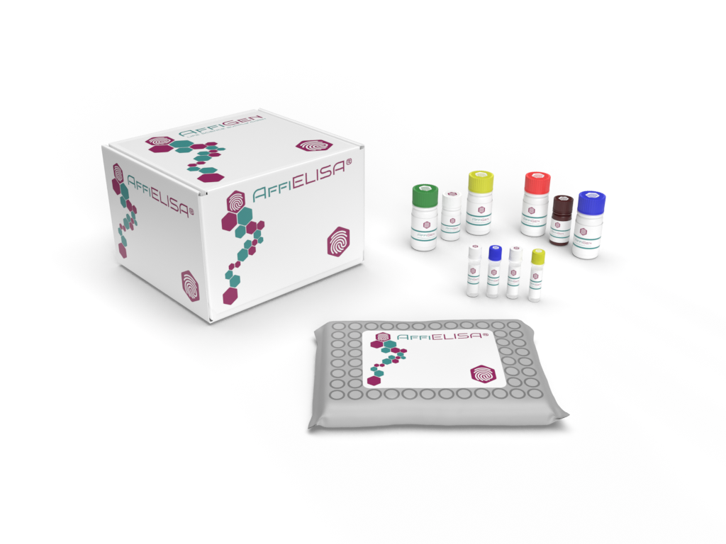
AffiELISA® Mouse IL6 Kit: High Sensitivity Interleukin 6 Microsample ELISA Detection

IL-6 Detection by ELISA: Core Principle and Workflow

Enzyme-Linked Immunosorbent Assay (ELISA) is a cornerstone in molecular and cellular biology for measuring cytokines like IL-6. The IL-6 ELISA Kit operates via a sandwich detection format, designed to capture and quantify IL-6 with exceptional accuracy.

Workflow Overview

  1. Coating Phase — A high-affinity monoclonal antibody specific to IL-6 is pre-coated onto a microtiter plate.

  2. Sample Incubation — Biological samples (serum, plasma, or culture supernatants) containing IL-6 are added to the wells for antigen–antibody binding.

  3. Detection Phase — A biotin-labeled secondary antibody binds to the captured IL-6 molecule.

  4. Signal Amplification — Streptavidin-HRP conjugate binds to biotin, catalyzing TMB substrate into a measurable chromogenic product.

  5. Quantification — The optical density (OD) is measured at 450 nm, and IL-6 concentration is determined using a standard curve.

More about ELISA principles and protocols can be found in resources from:

Technical Characteristics of IL-6 ELISA Kits

Parameter Specification
Detection Method Sandwich ELISA
Target Analyte Human IL-6 (Interleukin-6)
Sample Types Serum, Plasma, Cell Culture Supernatants
Sensitivity ≤ 0.5 pg/mL
Dynamic Range 1.5 pg/mL – 1000 pg/mL
Assay Time 2–3 hours
Detection Wavelength 450 nm
Standard Curve 7-Point Logarithmic Fit
Precision CV < 10 % (intra-assay)
Storage Temperature 2 – 8 °C
Regulatory Validation NIH, FDA, GLP-aligned

IL-6 ELISA kits are validated under Good Laboratory Practice (GLP) according to OECD guidelines and follow traceability standards referenced by the National Institute of Standards and Technology (NIST).

Applications of IL-6 Quantification in Experimental Biology

The IL-6 ELISA Kit is a critical analytical tool in multiple disciplines of life-science research. It provides quantitative insights into cytokine release, immune cell signaling, and biological stress responses.

1. Immunology and Cytokine Profiling

Researchers use IL-6 ELISA kits to assess cytokine dynamics in in vitro cell cultures, immunomodulation assays, and cytokine release studies.

2. Molecular Cell Biology

IL-6 concentration correlates with STAT3 activation and gene transcription under oxidative and metabolic stress.

3. Environmental and Stress Biology

Cytokine expression patterns, including IL-6, are studied to evaluate environmental stressors and pollutant exposure impacts on cultured cells.

4. Bioassay Development and Quality Control

Pharmaceutical and academic laboratories use IL-6 ELISA kits for calibration of cytokine standards, validation of recombinant protein production, and quality control of immunotherapeutic reagents.

Experimental Design and Data Analysis

Quantitative IL-6 data are analyzed by plotting the standard curve (OD 450 nm vs concentration) using 4-parameter logistic regression (4-PL). This ensures precise interpolation across the full dynamic range.

Key statistical principles are documented in:

Researchers typically perform each sample in duplicate or triplicate to minimize random variation and achieve robust coefficient of variation (CV) values under 10 %.

Interleukin-6 in Cytokine Networks

IL-6 does not function in isolation — it interacts with a network of cytokines including IL-1β, TNF-α, IL-8, and IL-10. Together, they orchestrate immune cell recruitment and acute-phase protein synthesis.

Cross-referenced cytokine data repositories:

Advantages of Using IL-6 ELISA Kits in Research

  • High Analytical Sensitivity: Detects picogram quantities of IL-6.

  • Exceptional Specificity: No cross-reactivity with structurally similar interleukins.

  • Wide Dynamic Range: Ideal for both low and high concentration samples.

  • Ease of Use: Pre-coated plates reduce procedural variability.

  • Batch-to-Batch Consistency: Ensured via strict quality control aligned with NIH Research Quality Assurance Standards.

These attributes make IL-6 ELISA kits indispensable for basic science research, protein quantification, immunochemistry, and reagent validation.

Optimization and Troubleshooting Guidelines

Common factors influencing assay performance include:

  • Improper washing — may increase background signal.

  • Incomplete incubation — can reduce sensitivity.

  • Incorrect standard dilution — may distort calibration curves.

  • Temperature variation — impacts enzyme kinetics and color development.

For best practices, refer to:

Quality Assurance and Storage

IL-6 ELISA reagents should be stored at 2–8 °C, protected from direct light. The microplates are typically sealed in aluminum pouches with desiccant to prevent humidity-induced degradation.

All reagents are tested for stability under accelerated aging and freeze-thaw stress following FDA stability testing protocols.

For academic users, additional validation information can be found in the NIH Reagent Validation Framework.

Integration with Multiplex Cytokine Platforms

Although the IL-6 ELISA Kit provides single-analyte detection, it is often integrated with multiplex assays such as Luminex® cytokine bead arrays for simultaneous quantification of IL-1β, IL-8, IL-10, TNF-α, and GM-CSF.

See additional references:

Data Interpretation in Research Context

When analyzing IL-6 concentrations, researchers often normalize data to total protein content or cell number. Relative quantification against housekeeping cytokines (e.g., IL-10) ensures reliable comparison between experimental groups.

Statistical frameworks for interpreting cytokine data are detailed at:

Why IL-6 ELISA Kits Are Essential Tools in Cytokine Research

The IL-6 ELISA platform offers:

  • Quantitative reproducibility

  • Automation compatibility for high-throughput workflows

  • Standardized calibration curves ensuring global reproducibility

  • Cross-platform comparability with proteomics and transcriptomic assays

It aligns with international assay validation guidelines ensuring data reliability, traceability, and compliance with institutional laboratory policies.

Summary

IL-6 ELISA Kits represent a gold standard for cytokine quantification in immunology and molecular biology. With unmatched precision, reproducibility, and sensitivity, they are critical for advancing our understanding of cytokine signaling, cellular responses, and molecular regulation.

By leveraging optimized assay design, robust antibodies, and high-purity standards, these kits deliver consistent, reproducible results suitable for use in academic, industrial, and government-funded laboratories worldwide.

High-performance IL-6 ELISA Kits for accurate measurement of Interleukin-6 in serum, plasma, and cell culture samples. Discover sensitivity ranges, workflow principles, and research applications. Trusted by NIH, CDC, and academic laboratories for immunology and protein quantification studies.