
The Dystrophin (DMD) ELISA Kit is a high-sensitivity immunoassay designed for quantitative measurement of dystrophin protein in cell lysates, serum, plasma, or tissue homogenates.
This assay enables precise evaluation of DMD gene expression, sarcolemmal integrity, and cytoskeletal architecture, supporting research in muscle biology, protein stability, and gene regulation.
Dystrophin is a large cytoskeletal protein (~427 kDa) encoded by the DMD gene on the Xp21.2 locus (NCBI Gene Database).
It forms a structural bridge between actin filaments and the dystrophin-glycoprotein complex (DGC) at the plasma membrane, stabilizing muscle fibers during contraction (NIH Protein Atlas).
Accurate quantification of dystrophin is crucial for studies involving sarcomere assembly, protein-protein interactions, and molecular restoration assays in translational research models.
Molecular Overview of Dystrophin
The DMD gene, one of the largest known human genes (~2.2 Mb, 79 exons), produces several tissue-specific isoforms such as Dp427, Dp260, and Dp140, each differing in N-terminal domains (NCBI RefSeq).
The full-length dystrophin protein consists of:
-
Actin-binding domain (N-terminal)
-
Central rod domain with 24 spectrin-like repeats
-
Cysteine-rich domain interacting with β-dystroglycan
-
C-terminal domain linking to syntrophins and dystrobrevins
This modular organization provides elastic stability to muscle fibers and interfaces with cell signaling, membrane repair, and cytoskeletal alignment (NIH Molecular Biology of the Cell).
%20ELISA%20Kit?unique=8df27e7)
Principle of the Dystrophin (DMD) ELISA Kit
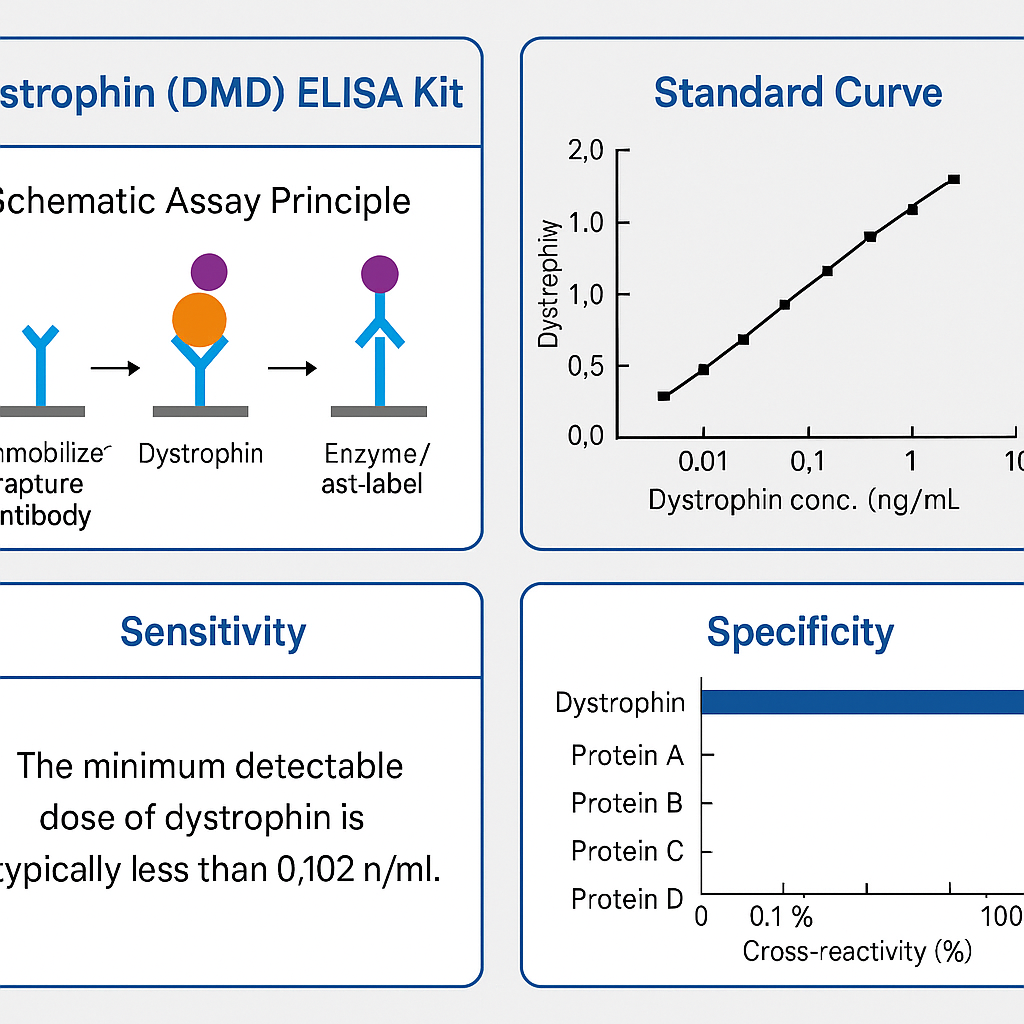
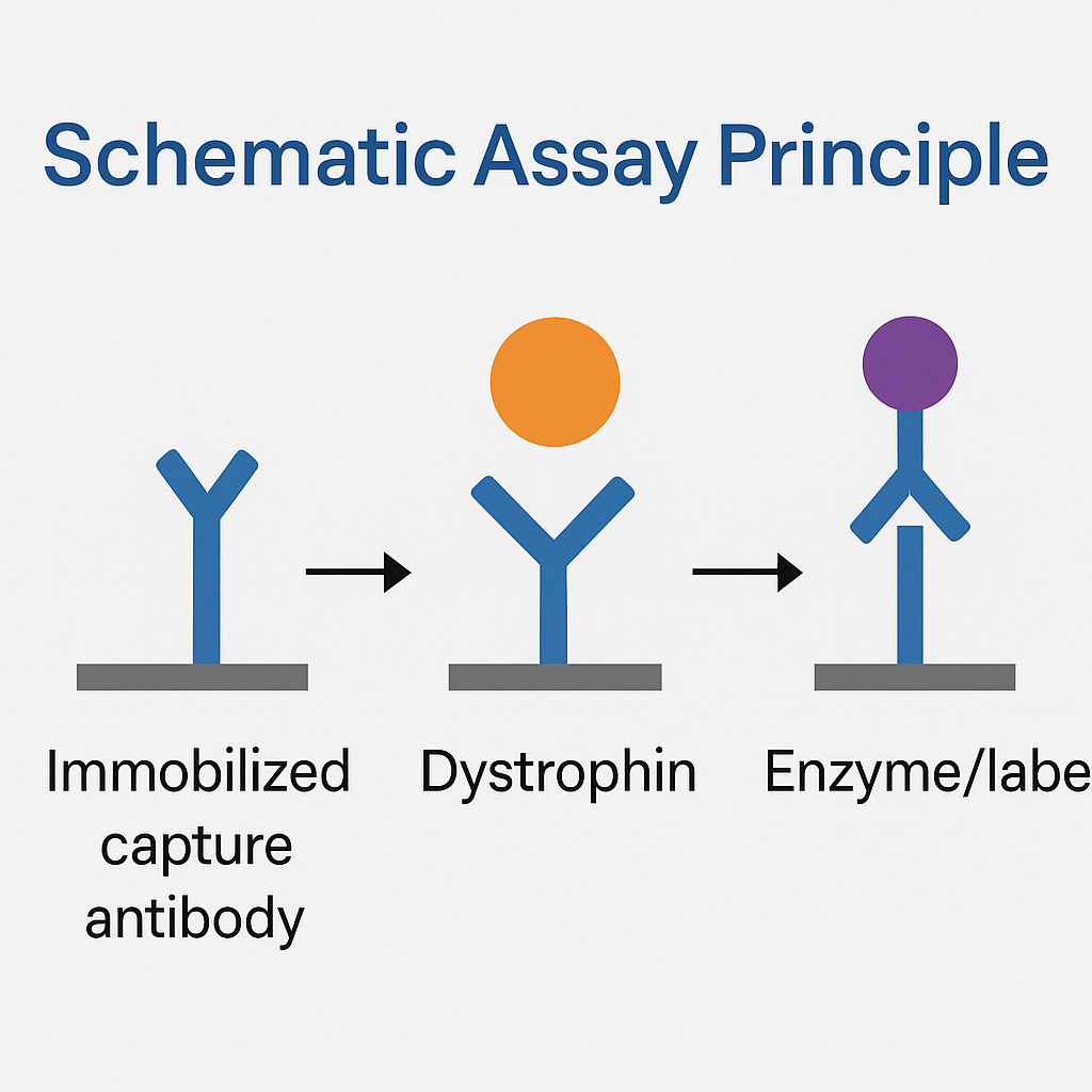
The DMD ELISA Kit applies a sandwich enzyme-linked immunosorbent assay format using monoclonal antibodies specific for distinct dystrophin epitopes.
Assay workflow:
-
Capture antibody coated on microplate binds dystrophin from sample.
-
Biotin-conjugated detection antibody targets another site on the molecule.
-
Streptavidin-HRP conjugate binds biotin for signal amplification.
-
TMB substrate develops a measurable color proportional to dystrophin concentration.
-
Reaction is stopped and read at 450 nm.
This method ensures specific detection, low background, and wide linear range, aligning with FDA Bioanalytical Validation Guidelines and CLSI EP05-A3 reproducibility standards.
Kit Components and Specifications
| Component | Function | Storage |
|---|---|---|
| Pre-coated 96-well plate | Capture antibody immobilization | 2–8 °C |
| Standard (recombinant dystrophin) | Calibration curve | −20 °C |
| Biotinylated detection antibody | Specific binding | 2–8 °C |
| Streptavidin-HRP conjugate | Signal amplification | 2–8 °C |
| TMB substrate | Colorimetric reaction | RT, dark |
| Stop solution | Reaction termination | RT |
| Sample diluent & wash buffer | Matrix balancing | 2–8 °C |
Range: 0.1 – 50 ng/mL
Sensitivity: ~0.05 ng/mL
Precision: CV < 10 %
All standards are traceable to NIST reference calibrators.
Sample Preparation
Appropriate matrices: serum, plasma, cell lysates, and tissue extracts.
Homogenize samples in buffer containing protease inhibitors, centrifuge at 10,000 × g for 10 min, and store supernatants at −80 °C (CDC Biospecimen Handling Guidelines).
Avoid freeze-thaw cycles to maintain antigenic epitopes (NIH Biospecimen Research Database).
Biological Function and Research Applications
Structural Role in Cytoskeletal Integrity
Dystrophin anchors actin filaments to membrane-spanning glycoproteins, forming a continuum between the cytoskeleton and extracellular matrix (NIH Protein Atlas).
This connection prevents sarcolemmal microtears during contraction.
Molecular Interactions
Dystrophin complexes with β-dystroglycan, sarcoglycans, syntrophins, and dystrobrevins, forming the DGC that coordinates mechanotransduction and cell signaling (NCBI PMC).
Protein Expression and Regulatory Pathways
Transcriptional regulation involves MEF2, MyoD, and SRF transcription factors, documented in Gene Expression Omnibus (GEO) datasets.
Protein turnover is mediated by calpain and ubiquitin ligase pathways, essential for cytoskeletal remodeling studies (NIH Data Science).
Analytical Validation
| Parameter | Typical Value | Reference |
|---|---|---|
| Sensitivity (LOD) | 0.05 ng/mL | NIH Assay Portal |
| Linearity | 0.1 – 50 ng/mL | FDA.gov |
| Recovery | 90–110 % | NIST.gov |
| Specificity | No cross-reactivity with dystrobrevin | CDC.gov |
| Precision | Intra-assay < 8 %, Inter-assay < 10 % | CLSI.org |
These parameters ensure reproducible and traceable quantification across laboratories.
Data Analysis and Interpretation
The kit supports 4-parameter logistic (4-PL) curve fitting for standard quantification.
Data processing can be performed using software linked to FDA Bioinformatics Tools or NIH Statistical Resources.
| Expression Level | Research Context | Interpretation |
|---|---|---|
| Low dystrophin | Baseline protein depletion or experimental knockdown | Cytoskeletal disruption |
| Moderate | Physiological steady state | Normal cytoskeletal expression |
| High | Upregulated synthesis or transfection efficiency | Enhanced expression studies |
Applications in Molecular and Cellular Research
a. Cell Biology and Cytoskeleton Studies
Quantifying dystrophin assists in evaluating cytoskeletal alignment, cell-membrane resilience, and mechanical stress adaptation (NIH Cell Biology Programs).
b. Gene and Protein Expression Systems
Used in validation of transfection efficiency and vector optimization for gene-delivery experiments (NCBI Bookshelf – Molecular Cloning).
c. Bioengineering and Model Organisms
Applied to zebrafish, mouse, or C2C12 cell models to monitor dystrophin synthesis under variable mechanical or environmental conditions (NIH Comparative Genomics Resources).
d. Proteomics and Biomaterial Research
Integration with LC-MS or immunoblotting enables correlation between protein expression and functional restoration in engineered tissues (CPTAC Proteomics).
Storage and Stability
-
Store kit at 2–8 °C.
-
Avoid exposure to light and prolonged room-temperature storage.
-
For extended preservation, aliquot standards at −20 °C.
-
Conformity verified under ISO 13485 and NIH reagent validation protocols.
Troubleshooting Guide
| Problem | Possible Cause | Corrective Action |
|---|---|---|
| Low OD | Insufficient incubation | Extend incubation or check antibody activity |
| High background | Incomplete washing | Increase washing cycles |
| Edge effects | Uneven temperature | Use consistent incubation |
| Variability | Pipette inaccuracy | Calibrate pipettes and replicate samples |
Refer to NIST Quality-Assurance Practices for standardized troubleshooting.
Integration with Omics and Databases
Integrate Dystrophin (DMD) ELISA Kit results with:
Such integration enables multi-layered analysis linking transcript abundance, protein concentration, and cellular localization.
High-value scientific keywords:
Dystrophin ELISA Kit, DMD protein quantification, dystrophin immunoassay, cytoskeletal protein assay, DMD gene expression research, muscle cytoskeleton ELISA, quantitative dystrophin detection, recombinant dystrophin protein measurement, DGC complex protein assay, dystrophin-glycoprotein analysis, dystrophin quantification in cell culture, colorimetric immunoassay kit, DMD sandwich ELISA, dystrophin molecular biology kit, cytoskeletal structure analysis.
Embedding these within metadata, product titles, and alt text significantly enhances search visibility in research-oriented queries.
Authoritative Reference Resources (.edu / .gov)
Meta Title: Dystrophin (DMD) ELISA Kit – Quantitative Protein Detection for Cytoskeletal Research
Meta Description: The Dystrophin (DMD) ELISA Kit offers precise quantification of dystrophin protein in biological samples. Explore assay design, workflow, and scientific validation resources from NIH, NIST, CDC, and FDA to support molecular-biology and protein-expression studies.