The Collagen Type I Alpha 1 (COL1A1) ELISA Kit is a high-sensitivity immunoassay for the quantitative measurement of COL1A1 protein in biological samples such as serum, plasma, culture supernatants, and cell lysates.
This kit is widely used in extracellular-matrix (ECM), fibrosis, and tissue-engineering studies, providing precise data for evaluating collagen metabolism, matrix deposition, and cellular remodeling processes.
Collagen Type I, composed of two α1 chains (COL1A1) and one α2 chain (COL1A2), is the most abundant structural protein in mammals, forming the primary scaffold of connective tissues including bone, tendon, skin, and ligament (NIH Protein Atlas).
Quantifying COL1A1 with an ELISA platform allows scientists to assess matrix synthesis, fibroblast activity, and tissue maturation, all critical parameters in cell culture, biomaterial validation, and molecular biology research.
Structural Biology of Collagen Type I Alpha 1
COL1A1 is encoded by the COL1A1 gene located on chromosome 17q21.33 (NCBI Gene Database).
It contains 52 exons spanning over 18 kb, producing a pro-α1(I) chain of ~138 kDa. Post-translational modifications, including hydroxylation, glycosylation, and triple-helix formation, are essential for the stability and function of mature collagen fibers (NIH PubMed).
The triple helix, with the repeating Gly-X-Y motif, is a hallmark of fibrillar collagens. Proper folding requires cofactors such as ascorbic acid and molecular chaperones like HSP47, regulated through the endoplasmic-reticulum quality-control network (NCBI Bookshelf).
%20%20ELISA%20Kit?unique=8df27e7)
Principle of the COL1A1 ELISA Assay
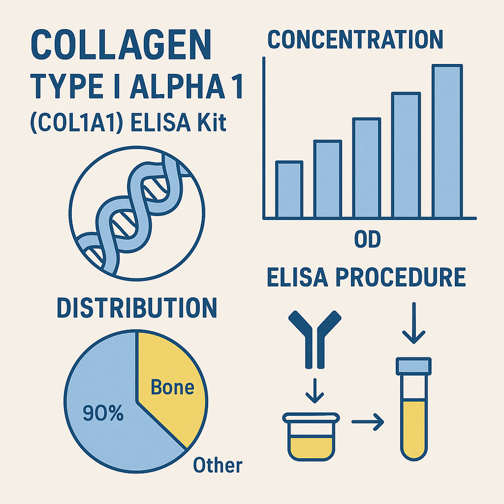
The Collagen Type I Alpha 1 ELISA Kit utilizes a sandwich enzyme-linked immunosorbent assay.
Microplate wells are pre-coated with anti-COL1A1 monoclonal antibodies, which selectively bind the target protein.
Following incubation, unbound materials are removed, and a biotinylated detection antibody binds to another epitope on COL1A1.
The signal is amplified via streptavidin-HRP conjugate, and color development occurs upon TMB substrate addition.
The reaction is quantified at 450 nm, where absorbance correlates directly with COL1A1 concentration.
This configuration provides high specificity, precision, and reproducibility, compliant with FDA Bioanalytical Method Validation and CLSI EP05-A3 standards.
Kit Components
| Component | Function | Storage |
|---|---|---|
| Pre-coated microplate | Capture antibody | 2–8 °C |
| Standard (recombinant COL1A1) | Calibration curve | −20 °C |
| Biotinylated detection antibody | Secondary binding | 2–8 °C |
| Streptavidin-HRP conjugate | Signal amplification | 2–8 °C |
| TMB substrate | Color development | RT, dark |
| Stop solution | Reaction termination | RT |
| Sample diluent, wash buffer | Matrix balancing | 2–8 °C |
Quantitative range: 0.05 – 10 ng/mL
Sensitivity: ~30 pg/mL
Precision: CV < 10 % intra-assay
All lots are traceable to NIST reference standards.
Sample Collection and Handling
Suitable specimens include human serum, EDTA plasma, or conditioned media.
Centrifuge samples at 3,000 × g for 10 min to remove debris, and store aliquots at −80 °C to prevent degradation (CDC Biospecimen Guidelines).
Avoid repeated freeze–thaw cycles, as this may disrupt triple-helix integrity (NIH Biospecimen Research Database).
Biological Role and Research Relevance
Extracellular Matrix Formation
COL1A1 forms the primary fibrillar framework of tissues. Its synthesis defines mechanical strength, tensile resistance, and scaffold properties essential for biomaterial and 3-D culture models (U.S. National Library of Medicine).
Cellular Signaling and Remodeling
Collagen I interacts with integrins (α1β1, α2β1), initiating FAK and MAPK signaling, thereby influencing cell proliferation and migration (NIH Signal Transduction Atlas).
Molecular Regulation
Transcriptional activation of COL1A1 involves factors like SP1, SMAD2/3, and TGF-β, as documented in Gene Expression Omnibus (GEO) datasets.
Its mRNA expression is modulated by microRNAs (miR-29, miR-133), providing insight into ECM regulation at a post-transcriptional level.
Assay Validation and Performance
| Parameter | Result | Reference |
|---|---|---|
| Sensitivity | 30 pg/mL | NIH Assay Portal |
| Specificity | No cross-reactivity with COL3A1/COL4A1 | FDA.gov |
| Recovery | 90–110 % | NIST.gov |
| Precision | CV < 10 % | CLSI.org |
| Dynamic Range | 0.05 – 10 ng/mL | CDC.gov |
These parameters ensure high reproducibility across multi-site experiments.
Data Interpretation
| COL1A1 Concentration | ECM Status | Possible Research Context |
|---|---|---|
| Low | Decreased matrix synthesis | Culture dedifferentiation, in vitro senescence |
| Normal | Baseline production | Homeostatic fibroblast culture |
| High | Active matrix remodeling | Tissue-engineering scaffold formation, growth-factor stimulation |
Data can be compared with expression metrics from the Human Protein Atlas or NIH CPTAC Proteomics.
Applications in Research Fields
Tissue Engineering
COL1A1 is used to evaluate biomaterial compatibility and matrix deposition during scaffold design (NIH NIBIB).
Regenerative Biology
COL1A1 quantification helps characterize stem-cell differentiation toward osteogenic or chondrogenic lineages (NIH Stem Cell Information).
Cell-Culture Optimization
Monitoring COL1A1 secretion informs culture condition optimization, mechanotransduction, and growth-factor influence (NCBI PMC).
Proteomic Profiling
Used in mass-spectrometry workflows and bioinformatics correlation with matrix genes from the UCSC Genome Browser.
Quality Control and Storage
Maintain all reagents at 2–8 °C, protecting TMB from light.
Standards can be aliquoted and stored at −20 °C for extended use.
Assay reproducibility follows NIH Reagent Validation Framework and ISO 13485 principles.
Troubleshooting Guide
| Issue | Probable Cause | Corrective Action |
|---|---|---|
| Low signal | Insufficient antigen binding | Extend incubation |
| High background | Incomplete washing | Increase wash cycles |
| Edge effects | Uneven temperature | Use uniform incubator |
| Inconsistent replicates | Pipette error | Calibrate instruments |
Refer to NIST Quality Assurance Protocols.
Integration with Omics Data
Researchers can align COL1A1 ELISA data with transcriptomic or proteomic datasets from:
This integration enhances interpretation in studies on matrix dynamics, mechanical biology, and protein folding.
Suggested Keywords:
Collagen Type I Alpha 1 ELISA Kit, COL1A1 protein quantification, extracellular matrix assay, collagen biosynthesis research, fibrillar collagen ELISA, structural protein analysis, ECM metabolism, recombinant collagen ELISA, quantitative immunoassay, collagen I alpha 1 measurement, in vitro matrix production, tissue engineering ELISA, COL1A1 protein detection, colorimetric ELISA kit, extracellular protein quantification.
Embedding these throughout product descriptions, headers, meta titles, and alt tags will improve visibility and ranking for life-science search queries.
Reference and Educational Resources (.edu / .gov)
Meta Title: Collagen Type I Alpha 1 (COL1A1) ELISA Kit – Quantitative Protein Assay for Matrix Research
Meta Description: Explore the Collagen Type I Alpha 1 (COL1A1) ELISA Kit for accurate quantification of extracellular-matrix proteins. Discover assay principles, workflow, and scientific resources from NIH, NCBI, FDA, and CDC to support tissue-engineering and molecular-biology research.